. 2002 Oct;76(20):10401-16.
doi: 10.1128/jvi.76.20.10401-10416.2002.

Life cycle heterogeneity in animal models of human papillomavirus-associated disease

Affiliations

Life cycle heterogeneity in animal models of human papillomavirus-associated disease

Woei Ling Peh et al. J Virol. 2002 Oct.

Abstract

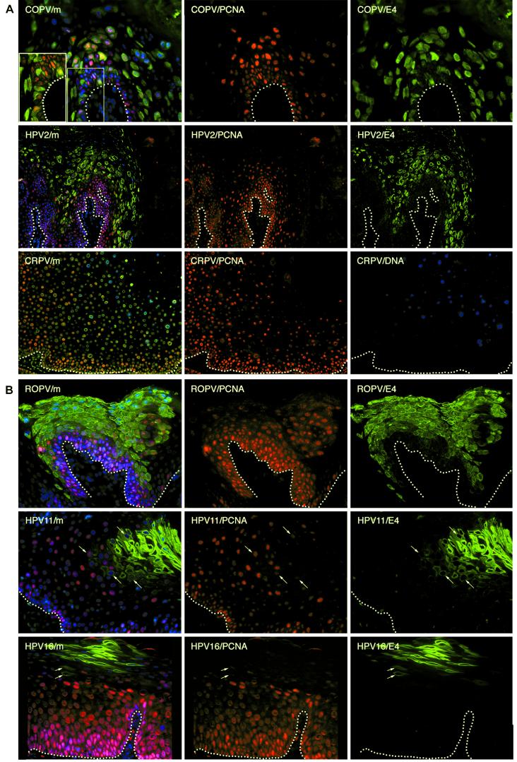
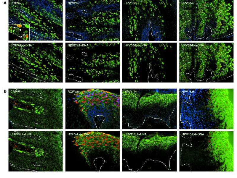
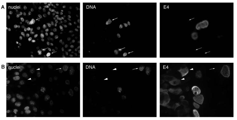
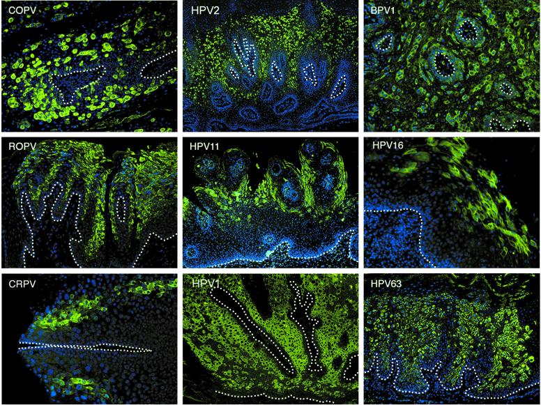
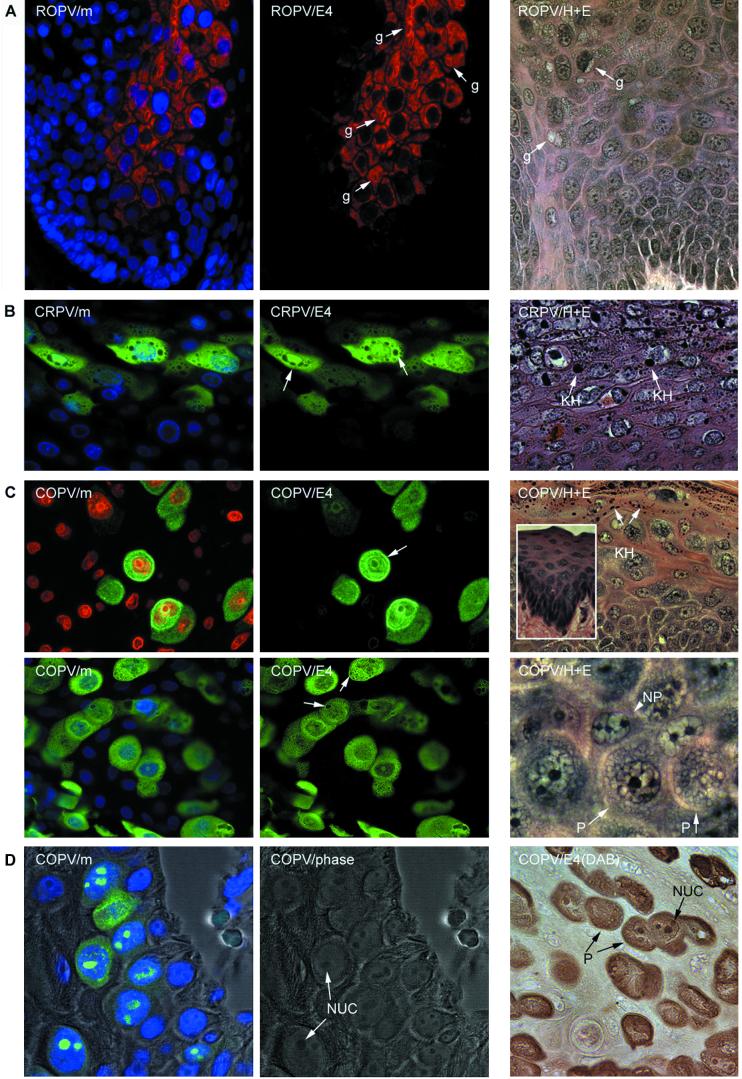
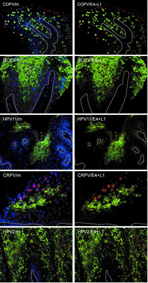
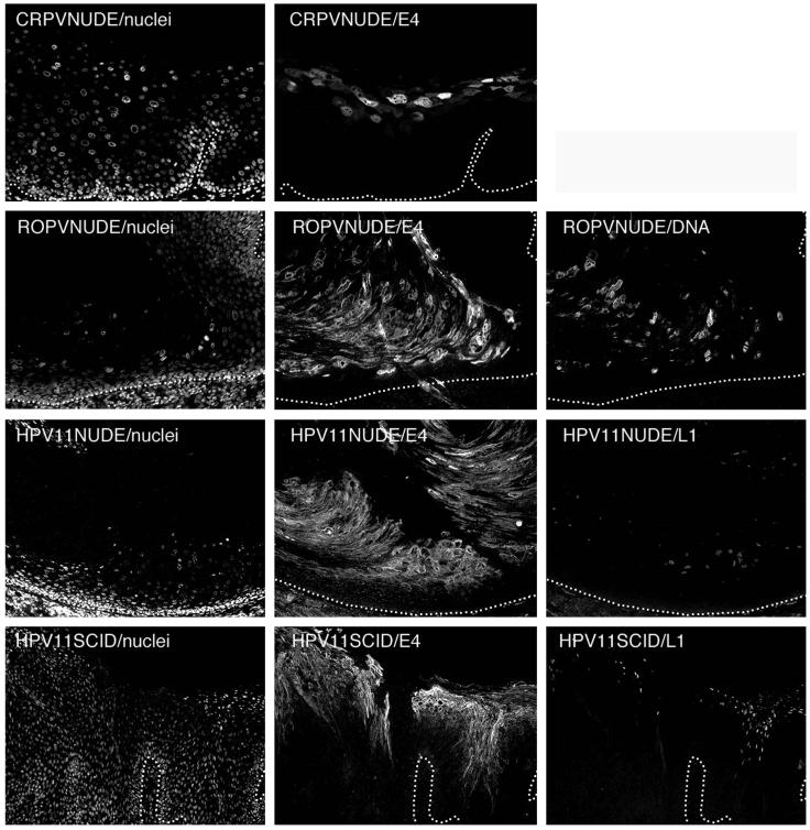
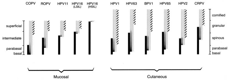
Animal papillomaviruses are widely used as models to study papillomavirus infection in humans despite differences in genome organization and tissue tropism. Here, we have investigated the extent to which animal models of papillomavirus infection resemble human disease by comparing the life cycles of 10 different papillomavirus types. Three phases in the life cycles of all viruses were apparent using antibodies that distinguish between early events, the onset of viral genome amplification, and the expression of capsid proteins. The initiation of these phases follows a highly ordered pattern that appears important for the production of virus particles. The viruses examined included canine oral papillomavirus, rabbit oral papillomavirus (ROPV), cottontail rabbit papillomavirus (CRPV), bovine papillomavirus type 1, and human papillomavirus types 1, 2, 11, and 16. Each papillomavirus type showed a distinctive gene expression pattern that could be explained in part by differences in tissue tropism, transmission route, and persistence. As the timing of life cycle events affects the accessibility of viral antigens to the immune system, the ideal model system should resemble human mucosal infection if vaccine design is to be effective. Of the model systems examined here, only ROPV had a tissue tropism and a life cycle organization that resembled those of the human mucosal types. ROPV appears most appropriate for studies of the life cycles of mucosal papillomavirus types and for the development of prophylactic vaccines. The persistence of abortive infections caused by CRPV offers advantages for the development of therapeutic vaccines.


Figures

FIG.1.
FIG.1.
Heterogeneity in the timing of events during the life cycles of human and animal papillomaviruses. (A) Tissue sections from a mucosal lesion caused by COPV (top row) and a cutaneous lesion caused by HPV2 (middle row) were double stained using antibodies to E4 (green) andPCNA (red) before being counterstained with DAPI (blue) to visualize cell nuclei. Surrogate markers of E7 expression (PCNA) were confined to sporadic cells in the lowest parabasal layers. E4 (green) was first detected in these cells as they migrated through the lower layers of the epithelium. E4 was expressed in the basal layer in lesions caused by COPV and in a few cells above the basal layer in lesions caused by HPV2. Surrogate markers of E7 were lost soon after the appearance of E4. The merged images (m) are shown at the left. A section through a cutaneous lesion caused by CRPV is shown in the bottom row after immunostaining to detect PCNA (red) and in situ hybridization to detect viral genome amplification (blue). The nuclei were counterstained using Sytox green. Surrogate markers of E7 expression (PCNA) did not persist to the epithelial surface and were lost soon after the onset of viral genome amplification (blue). Genome amplification began in the mid-spinous layers in lesions caused by CRPV. The broken lines indicate the positions of the basal layers. The images were taken using a 10× (COPV and HPV2) or 20× (COPV/m inset) objective. (B) Tissue sections from mucosal lesions caused by ROPV (top row), HPV11 (middle row), and HPV16 (bottom row) were double stained with antibodies to E4 (green) and PCNA (red) before being counterstained with DAPI (blue) to visualize cell nuclei. The merged images are shown at the left. In such lesions, surrogate markers of E7 expression (PCNA; red) usually persisted into the upper epithelial layers but were lost following the expression of E4. E4 was rarely detected in the lower epithelial layers in lesions caused by HPV11 and -16. Cells expressing both E4 and PCNA are indicated by arrows in the HPV11 and HPV16 images. The broken lines indicate the positions of the basal layers. The images were taken using a 20× objective.
FIG.2.
FIG.2.
The onset of vegetative viral genome amplification coincides closely with the expression of E4 in all papillomavirus infections. (A) Tissue sections from a mucosal lesion caused by COPV and cutaneous lesions caused by HPV2, HPV65, and HPV63 were probed to detect the presence of amplified viral DNA (red) and E4 (green). The nuclei were counterstained using DAPI (blue). Genome amplification was generally confined to cells expressing E4 (green). In warts caused by COPV, HPV2, and HPV65, such cells were distributed sporadically throughout the lesion. The broken lines indicate the positions of the basal layers. The images were taken using a 10× objective. The inset shows COPV E4 and viral genome amplification in the basal layer (green, E4; red, amplified viral DNA; blue, DAPI counterstain). The images were taken using a 40× objective. (B) Tissue section from a cutaneous lesion caused by CRPV and from mucosal lesions caused by ROPV, HPV11, and HPV16 were probed by in situ hybridization to detect cells that had amplified viral DNA (red). E4 was detected by using specific antibodies to the E4 proteins of each virus type (green), and the nuclei were counterstained using DAPI (blue). E4 expression and viral genome amplification were restricted to a band of cells in the middle and upper epithelial layers. The broken lines indicate the positions of the basal layers. The images were taken using a 10× objective.
FIG. 3.
FIG. 3.
E4 expression and the onset of genome amplification are not necessarily coincident. (A and B) Tissue sections taken from two different COPV-infected oral lesions. Tissue sections were double stained for the presence of viral genome amplification (DNA) and for E4 expression (E4). Viral DNA was occasionally detected in the absence of E4 (cells indicated by arrows). The nuclei were counterstained using Sytox green (nuclei). (B) E4 expression in cells (indicated by arrowheads) that are not supporting viral genome amplification. A cell (indicated by an arrow) supporting viral genome amplification but lacking E4 is shown in the same field of view. The images were taken using a 40× objective.
FIG. 4.
FIG. 4.
Timing of late events is highly conserved among lesions caused by the same papillomavirus type. Similarities in the patterns of E4 expression and genome amplification confirmed that E4 was an effective marker of the late stages of the papillomavirus life cycle. Tissue biopsies infected by different papillomaviruses were sectioned and stained using E4-specific antibodies (green) before being counterstained with DAPI (blue). The staining patterns illustrated were typical of those seen following the examination of five or more independent biopsy specimens infected by each virus type (except for HPV63, for which three lesions were examined [see Materials and Methods]). The timing of late-stage onset was a characteristic feature of each infecting virus type. All images were taken using a 4× objective except for the HPV16 and CRPV images, which were taken using a 10× objective lens. The basal layers are indicated by the broken lines.
FIG.5.
FIG.5.
Intracellular distribution of the E4 proteins of animal papillomaviruses. (A) The E4 protein of ROPV (red) is predominantly cytoplasmic and is associated with inclusion granules (center image, g) similar to those seen in cutaneous lesions caused by HPV1. The nuclei werecounterstained with DAPI (blue) and are visible in the merged image at the left (ROPV/m). The E4 granules (g) are also shown in the hematoxylin- and eosin-stained image at the right (labeled ROPV/H+E). The images were taken using a 40× objective. (B) The E4 protein of CRPV (green) is distributed throughout the nucleus and the cytoplasm. The nuclei were counterstained with DAPI (blue) and are visible in the merged image shown on the left (CRPV/m). The cytoplasmic structures that did not stain with antibodies to E4 are keratohyalin granules (center image, arrows). They are also shown (KH) in the hematoxylin- and eosin-stained image (CRPV/H+E) on the right. (C) The E4 protein of COPV (green) was cytoplasmic and nuclear but was also associated with the nuclear and cellular periphery (upper center image, arrow). The nuclei were counterstained with either propidium iodide (upper left image, red) or DAPI (lower left image, blue). A granular pattern was apparent in the cytoplasm of some cells (granule-like structures [arrows] in lower center image). Keratohyalin granules (KH) are abundant in COPV-induced warts and are shown in the hematoxylin- and eosin-stained image (COPV/H+E; upper right). The surrounding mucosal epithelium is devoid of keratohyalin (inset). Cells expressing COPV E4 had a characteristic morphology that may result from the presence of E4 inclusion granules. The permissive cells (P) that express E4 and the nonpermissive cells (NP) that do not express E4 are shown in the hematoxylin- and eosin-stained image on the lower right. (D) In the lower epithelial layers of experimental warts caused by COPV, nuclear E4 protein (green) was found associated with the nucleoli. At the left (COPV/m), the E4 and DAPI (blue) stain is shown as an overlay of the phase-contrast image. The phase-contrast image is shown in the center panel to indicate the presence of the nucleoli (NUC). The presence of the permissive “granular” cells (P) and the nucleoli is clearly visualized in the immunoperoxidase stain (brown) shown on the right [COPV/E4 (DAB)].
FIG. 6.
FIG. 6.
Expression of capsid proteins follows expression of E4 in lesions caused by different papillomavirus types. Tissue sections of lesions caused by COPV, ROPV, HPV11, CRPV, and HPV2 were double stained using antibodies to the L1 capsid protein (red) and E4 (green) before being counterstained with DAPI (blue). The merged images (m) are shown on the left. Although E4 expression always precedes the expression of L1, the distance between the first appearance of E4 and the first appearance of L1 varied considerably. The positions of the basal layers are indicated by broken lines. The images were taken using a 10× (HPV2, HPV11, and COPV) or 20× (CRPV) objective.
FIG. 7.
FIG. 7.
The timing of late gene expression in xenografts resembles that seen during natural infection. Epithelial tissue infected by CRPV or ROPV was propagated under the kidney capsule of nude mice (images labeled CRPVNUDE and ROPVNUDE) before being stained for E4 and (for ROPV) genome amplification. The nuclei were counterstained with DAPI and are shown on the left. In all cases, the timing of late-stage activation (as determined by E4 expression) was similar to that seen following natural and experimental infection of the natural host (Fig. 4). Genome amplification and E4 expression coincided closely (images labeled ROPVNUDE/DNA and ROPVNUDE/E4). Epithelial tissue infected by HPV11 was propagated under the kidney capsule of nude mice or as a skin graft in SCID mice. L1 expression followed that of E4 in renal capsule xenografts infected with HPV11 (images labeled HPV11NUDE/E4 and HPV11NUDE/L1). This pattern of expression was also seen in skin grafts propagated on SCID mice (HPV11SCID/E4 and HPV11SCID/L1). The morphology of the skin xenografts closely resembled that seen in HPV11-induced genital lesions (Fig. 2 and 4). The images were taken using a 10× (ROPV and HPV11) or 20× objective. The broken lines indicate the positions of the epithelial basal layers.
FIG. 8.
FIG. 8.
The regulation of early and late events in lesions caused by different papillomavirus types. The timing of life cycle events in lesions caused by animal and human papillomaviruses is indicated by the bars. The shaded bars show the presence of amplified viral DNA, while the stippled bars show the extent of E4 expression. The expression pattern of surrogate markers of E7 is shown by the solid bars, whereas the L1 expression pattern is indicated by the hatched bars. The darker region at the bottom of the shaded bars indicates the region where vegetative viral genome amplification is thought to occur. Mucosal epithelial tissue infected by COPV, ROPV, HPV11, and HPV16 is divided into four layers, shown on the left. Cutaneous tissue infected by HPV1, HPV63, BPV1, HPV65, HPV2, and CRPV is divided into five layers, shown on the right. For each group of papillomaviruses, those that initiate their late events in the lower epithelial layers are shown on the left. Viruses that initiate their late events close to the epithelial surface are shown on the right. COPV triggers late events in the lowest epithelial layers. Lesions caused by HPV11 and HPV16 (LSIL) usually support late gene expression only in the upper half of the epidermis, whereas late events are not always supported in HSIL. In all instances, the loss of surrogate markers of E7 does not occur until after E4 has accumulated to detectable levels.

References

    1. Alderborn, A., and S. Burnett. 1994. Regulation of DNA synthesis in division-arrested mouse C127 cells permissive for bovine papillomavirus DNA amplification. J. Virol. 68:4349-4357. - PMC - PubMed
    1. Ashmole, I., P. H. Gallimore, and S. Roberts. 1998. Identification of conserved hydrophobic C-terminal residues of the human papillomavirus type 1 E1E4 protein necessary for E4 oligomerisation in vivo. Virology 240:221-231. - PubMed
    1. Baker, C. C., and P. M. Howley. 1987. Differential promoter utilization by the bovine papillomavirus in transformed cells and in productively infected wart tissues. EMBO J. 6:1027-1035. - PMC - PubMed
    1. Barrera-Oro, J. G., K. O. Smith, and J. L. Melnick. 1962. Quantitation of papova virus particles in human warts. J. Natl. Cancer Inst. 29:583-595. - PubMed
    1. Bell, J. A., J. P. Sundberg, S. J. Ghim, J. Newsome, A. B. Jenson, and R. Schlegel. 1994. A formalin-inactivated vaccine protects against mucosal papillomavirus infection: a canine model. Pathobiology 62:194-198. - PubMed

Publication types

MeSH terms

Substances