. 2002 Oct;76(20):10455-64.
doi: 10.1128/jvi.76.20.10455-10464.2002.

Influenza virus can enter and infect cells in the absence of clathrin-mediated endocytosis

Affiliations

Influenza virus can enter and infect cells in the absence of clathrin-mediated endocytosis

Sara B Sieczkarski et al. J Virol. 2002 Oct.

Abstract

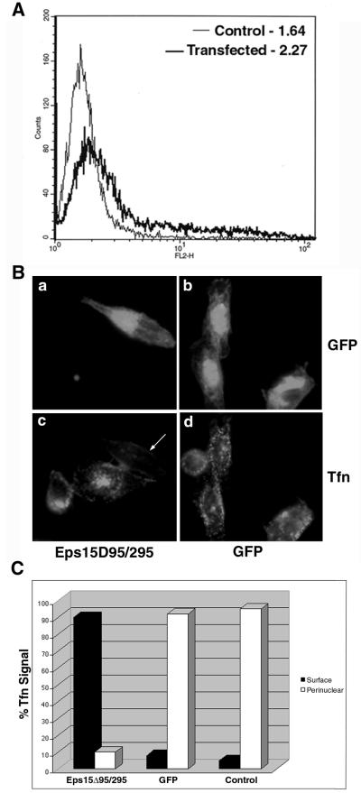
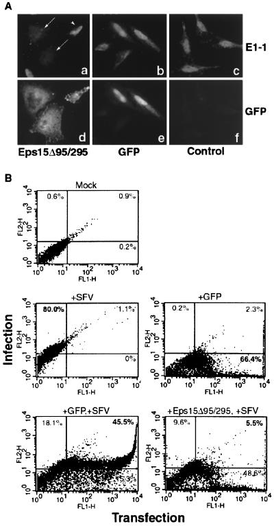
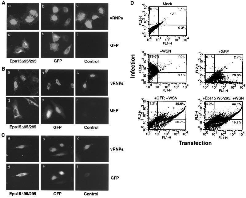
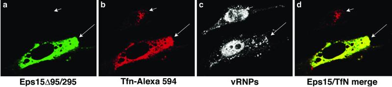
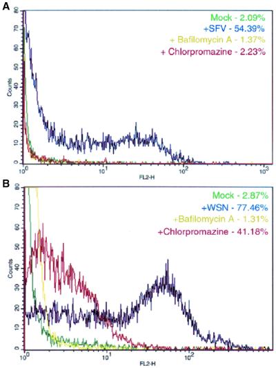
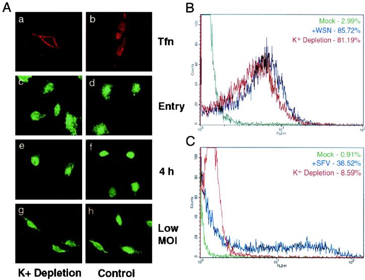
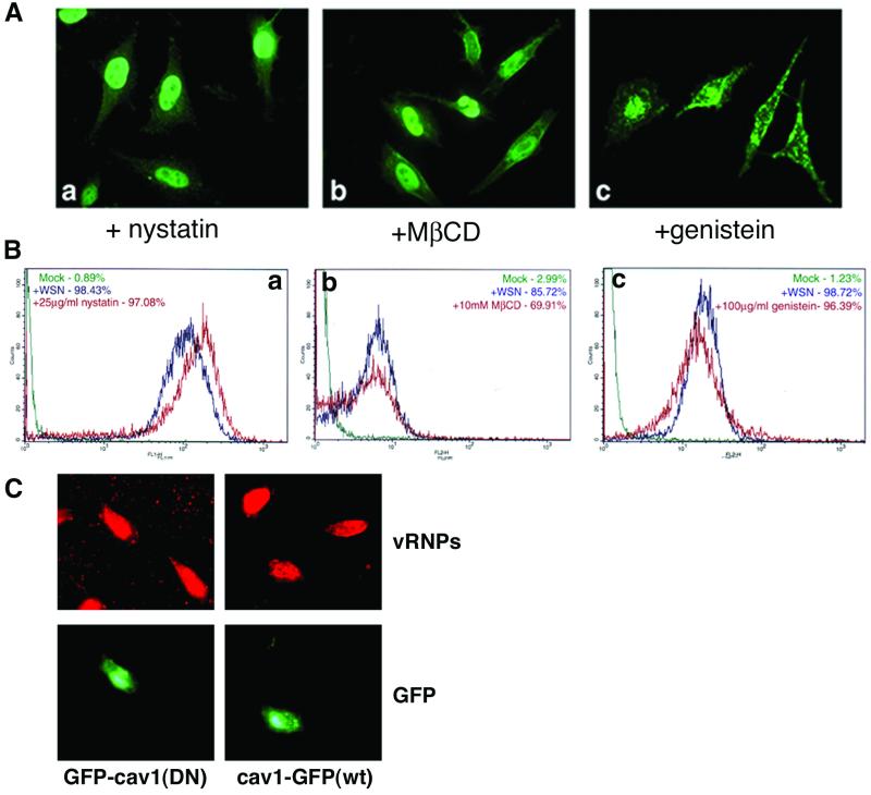
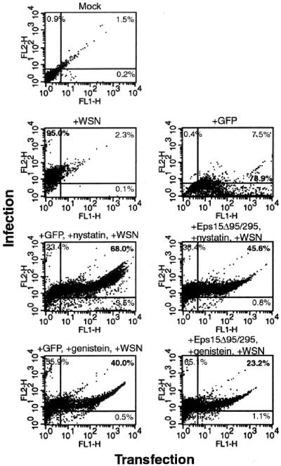
Influenza virus has been described to enter host cells via clathrin-mediated endocytosis. However, it has also been suggested that other endocytic routes may provide additional entry pathways. Here we show that influenza virus may enter and infect HeLa cells that are unable to take up ligands by clathrin-mediated endocytosis. By overexpressing a dominant-negative form of the Eps15 protein to inhibit clathrin-mediated endocytosis, we demonstrate that while transferrin uptake and Semliki Forest virus infection were prevented, influenza virus could enter and infect cells expressing Eps15Delta95/295. This finding is supported by the successful infection of cells with influenza virus in the presence of chemical treatments that block endocytosis, namely, chlorpromazine and potassium depletion. We show also that influenza virus may infect cells incapable of uptake by caveolae. Treatment with the inhibitors nystatin, methyl-beta-cyclodextrin, and genistein, as well as transfection of cells with dominant-negative caveolin-1, had no effect on influenza virus infection. By combining inhibitory methods to block both clathrin-mediated endocytosis and uptake by caveolae in the same cell, we demonstrate that influenza virus may infect cells by an additional non-clathrin-dependent, non-caveola-dependent endocytic pathway. We believe this to be the first conclusive analysis of virus entry via such a non-clathrin-dependent pathway, in addition to the traditional clathrin-dependent route.


Figures

FIG. 1.
FIG. 1.
GFP-Eps15Δ95/295 is overexpressed and prevents transferrin uptake. (A) HeLa cells were either transiently transfected with GFP-Eps15Δ95/295 or left as untransfected controls. Cells were prepared for FACS analysis by using the monoclonal antibody against Eps15 and an Alexa 568-labeled secondary antibody. Shaded curve, untransfected cells; solid curve, transfected cells. Numbers indicate median fluorescence expression as determined by FluoView software. At least 10,000 cells were counted. (B) HeLa cells were transiently transfected with either GFP-Eps15Δ95/295 or GFP alone. Alexa 594-labeled transferrin (Tfn) was bound to serum-starved cells for 20 min at 4°C. Cells were washed, transferred to 37°C for 15 min, washed with low-pH glycine to remove uninternalized ligand, and fixed for viewing. (C) Transferrin uptake quantitated by indirect immunofluorescence. One hundred cells from each category were counted, and residual surface transferrin was distinguished from a strong perinuclear signal.
FIG. 2.
FIG. 2.
SFV infection is prevented in GFP-Eps15Δ95/295-expressing cells. HeLa cells either were transiently transfected with either GFP or GFP-Eps15Δ95/295 or were left as untransfected controls before 5 h of infection. Infection was monitored using monoclonal antibody E1-1. (A) Indirect immunofluorescence monitoring of infection. Arrows indicate uninfected cells; arrowhead indicates a transfected, infected cell. (B) FACS analysis to quantitate immunofluorescence results. Infection is monitored on the y axis, and transfection is monitored on the x axis. The percentage of cells within each quadrant is given.
FIG. 3.
FIG. 3.
GFP-Eps15Δ95/295 expression does not prevent influenza virus infection. HeLa cells either were transiently transfected with either GFP or GFP-Eps15Δ95/295 or were left as untransfected controls before infection with influenza virus. Infection was monitored using monoclonal antibody H10, L16-4R5 against NP. (A) Entry assay. Cells were infected with ∼100 to 200 PFU per cell, and the virus was allowed to internalize for 60 min at 37°C. (B) Infection. Cells were infected with ∼1 to 5 PFU per cell and then incubated at 37°C for 4 h. (C) Low-MOI infection. Cells were infected with ∼0.1 PFU per cell and incubated at 37°C for 4 h. (D) FACS analysis of infection assay. Infection is monitored on the y axis, and transfection is monitored on the x axis. The percentage of cells within each quadrant is given.
FIG. 4.
FIG. 4.
Confocal microscopy of GFP-Eps15Δ95/295-expressing cells with influenza virus and transferrin (Tfn). HeLa cells were transiently transfected with GFP-Eps15Δ95/295 before high-MOI infection with influenza virus. The transferrin uptake assay was then performed; note that the cells were not acid washed after incubation. Influenza virus localization was determined by indirect immunofluorescence with an anti-NP monoclonal antibody. Images were created using Fluoview software and were transferred into Adobe Photoshop. The short arrow in panel b and the long arrow in panel c indicate transferrin and influenza vRNP signal localization, respectively, in untransfected cells.
FIG. 5.
FIG. 5.
Chlorpromazine treatment does not inhibit influenza virus infection. (A) HeLa cells were infected with SFV for 5 h, and infection was monitored using monoclonal antibody E1-1. (B) HeLa cells were infected with influenza virus for 4 h, and infection was monitored using an anti-NP monoclonal antibody. Cells were treated with either 25 nM bafilomycin A or 10 μg of chlorpromazine/ml during both the adsorption and incubation periods. Infection was monitored by FACS analysis. Key indicates treatment of cells. Background was determined by residual antibody staining on uninfected cells.
FIG. 6.
FIG. 6.
K+ depletion does not inhibit influenza virus infection. (A) HeLa cells were shocked in a hyptonic medium for 5 min before either an influenza vRNP entry assay, a 4-h infection (1 PFU/cell), a low-MOI infection (<0.1 PFU/cell), or a transferrin (Tfn) uptake assay in a K+-free medium. Influenza virus infection was detected by indirect immunofluorescence with a monoclonal antibody against NP. Note that cells were not acid washed after transferrin uptake. (B) FACS analysis. HeLa cells were shocked in a hyptonic medium for 5 min before an influenza virus entry assay in a K+-free medium. Influenza virus was detected by a monoclonal antibody against NP. (C) FACS analysis of SFV infection in cells depleted of potassium. HeLa cells were shocked in a hypotonic medium for 5 min before 5 h of infection with SFV in a K+-free medium. Virus infection was detected by monoclonal antibody E1-1.
FIG. 7.
FIG. 7.
Treatment with caveola-inhibiting drugs does not prevent influenza virus infection. HeLa cells were treated with either 25 μg of nystatin/ml, 10 mM MβCD, or 100 μg of genistein/ml during both the adsorption and incubation periods of influenza virus infection. Influenza virus infection was detected with a monoclonal antibody against NP. (A) Indirect immunofluorescence analysis of 4-h influenza virus infection in drug-treated cells. (B) FACS analysis of entry assay infection in drug-treated cells. Key indicates treatment of cells. Background was determined by residual antibody staining on uninfected cells. (C) HeLa cells were transiently transfected with wild-type or dominant-negative caveolin-1 constructs before entry assay infection with influenza virus. Transfected cells are indicated by GFP expression; infection was monitored via indirect immunofluorescence using a monoclonal antibody against NP.
FIG. 8.
FIG. 8.
Inhibition of both clathrin-mediated endocytosis and uptake by caveolae does not prevent influenza virus infection. HeLa cells were transiently transfected with either GFP or GFP-Eps15Δ95/295. Cells were treated with either 25 μg of nystatin/ml or 100 μg of genistein/ml during a high-MOI infection with influenza virus. Infection was monitored by FACS analysis using a monoclonal antibody against NP. Infection is monitored on the y axis, and transfection is monitored on the x axis. The percentage of cells within each quadrant is given.

Similar articles

Cited by

References

    1. Ahn, A., D. L. Gibbons, and M. Kielian. 2002. The fusion peptide of Semliki Forest virus associates with sterol-rich membrane domains. J. Virol. 76:3267-3275. - PMC - PubMed
    1. Bayer, N., D. Schober, M. Huttinger, D. Blaas, and R. Fuchs. 2001. Inhibition of clathrin-dependent endocytosis has multiple effects on human rhinovirus serotype 2 cell entry. J. Biol. Chem. 276:3952-3962. - PubMed
    1. Benmerah, A., M. Bayrou, N. Cerf-Bensussan, and A. Dautry-Varsat. 1999. Inhibition of clathrin-coated pit assembly by an Eps15 mutant. J. Cell Sci. 112:1303-1311. - PubMed
    1. Benmerah, A., B. Begue, A. Dautry-Varsat, and N. Cerf-Bensussan. 1996. The ear of alpha-adaptin interacts with the COOH-terminal domain of the Eps 15 protein. J. Biol. Chem. 271:12111-12116. - PubMed
    1. Benmerah, A., J. Gagnon, B. Begue, B. Megarbane, A. Dautry-Varsat, and N. Cerf-Bensussan. 1995. The tyrosine kinase substrate eps15 is constitutively associated with the plasma membrane adaptor AP-2. J. Cell Biol. 131:1831-1838. - PMC - PubMed

Publication types

MeSH terms

LinkOut - more resources