. 2002 Oct;76(20):10540-5.
doi: 10.1128/jvi.76.20.10540-10545.2002.

Protective antiviral immune responses to pseudorabies virus induced by DNA vaccination using dimethyldioctadecylammonium bromide as an adjuvant

Affiliations

Protective antiviral immune responses to pseudorabies virus induced by DNA vaccination using dimethyldioctadecylammonium bromide as an adjuvant

Eugene M A van Rooij et al. J Virol. 2002 Oct.

Abstract

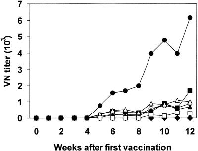
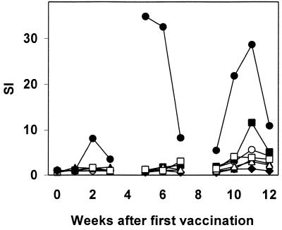
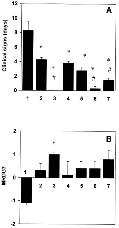
To enhance the efficacy of a DNA vaccine against pseudorabies virus (PRV), we evaluated the adjuvant properties of plasmids coding for gamma interferon or interleukin-12, of CpG immunostimulatory motifs, and of the conventional adjuvants dimethyldioctadecylammonium bromide in water (DDA) and sulfolipo-cyclodextrin in squalene in water. We demonstrate that a DNA vaccine combined with DDA, but not with the other adjuvants, induced significantly stronger immune responses than plasmid vaccination alone. Moreover, pigs vaccinated in the presence of DDA were protected against clinical disease and shed significantly less PRV after challenge infection. This is the first study to demonstrate that DDA, a conventional adjuvant, enhances DNA vaccine-induced antiviral immunity.


Figures

FIG. 1.
FIG. 1.
Expression of biologically active pIL12 by transfected COS-7 cells. Culture medium samples of COS-7 cells transfected with the plasmid VR1012 (1), VR-p40 (2), or VR-pIL12 (3) were collected 72 h posttransfection, and the ability to stimulate the proliferation of human PBMCs was evaluated. Culture medium (4) and medium containing 12 ng of recombinant human IL-12/ml (5) were used as controls. Data are expressed as the geometric means + the standard errors of the means. Results shown are representative of two similar experiments.
FIG. 2.
FIG. 2.
Expression of biologically active pIFN-γ by transfected COS-7 cells. Culture medium samples of COS-7 cells transfected with VR1012 (white bars) or VR1012-p IFN-γ (black bars) were collected 72 h posttransfection, diluted 20, 200, 2,000, or 20,000 times, and assayed for antiviral activity, using an FMDV plaque reduction bioassay. Data are expressed as the geometric means + the standard errors of the means. Results shown are representative of two similar experiments.
FIG. 3.
FIG. 3.
Analysis of PRV-neutralizing antibodies in sera of immunized pigs. Pigs were vaccinated at weeks 0, 4, and 8 with VR1012 (♦), VR-gB + VR-gD (▴), VR-gB + VR-gD with pUC 18 (▵), VR-IFN-γ (▪), VR-pIL12 (○), SL-CD (□), or DDA (•). Samples from the individual pigs were tested. Data are expressed as geometric mean titers of the different groups. Differences in group averages were tested for statistical significance by a parametric one-way analysis of variance (ANOVA) (95% significance level) for the entire observation period after each vaccination and not for the individual time points. For reasons of clarity, error bars are not shown.
FIG. 4.
FIG. 4.
Induction of PRV-specific T-cell responses in vaccinated pigs. Pigs were vaccinated at weeks 0, 4, and 8 with VR1012 (♦), VR-gB + VR-gD (▴), VR-gB + VR-gD with pUC 18 (▵), VR-p IFN-γ (▪), VR-pIL12 (○), SL-CD (□), or DDA (•). PBMCs were stimulated for 4 days with medium or live PRV, after which [3H]thymidine incorporation levels were determined. Data are expressed as SI values. Based on the SI values of the control group (mean + 3× standard deviation), an SI of ≥2.5 was considered positive. Throughout the experiments, counts of mock-stimulated PBMCs ranged from 300 to 1,500 and standard errors of the means of quadruplicates were less than 20%. Samples from the individual pigs were tested. Data are expressed as geometric mean SI values of the different groups. Differences in group averages were tested for statistical significance by a parametric one-way ANOVA (95% significance level) for the entire observation period after each vaccination and not for the individual time points. For reasons of clarity, error bars are not shown.
FIG. 5.
FIG. 5.
Virus excretion after challenge infection with PRV strain NIA-3. (A) Pigs vaccinated with VR1012 (♦), VR-gB + VR-gD (▴), VR-gB + VR-gD with pUC 18 (▵), VR-p IFN-γ (▪), VR-pIL12 (○), SL-CD (□), or DDA (•) were challenge infected 6 weeks after the third vaccination. Samples from the individual pigs were tested. Data are expressed as geometric mean virus titers (log10) per gram of oropharyngeal fluid for the different groups. Differences in group averages were tested for statistical significance by a parametric one-way ANOVA (95% significance level). For reasons of clarity, error bars are not shown. (B) Pigs vaccinated with VR1012 (♦) or VR-IE with DDA (⋄) were challenge infected 6 weeks after the third vaccination. Samples from the individual pigs were tested. Data are expressed as geometric mean virus titers (log10) per gram of oropharyngeal fluid for the different groups. There were no significant differences in results between VR-IE- and sham (VR1012)-vaccinated pigs.
FIG. 6.
FIG. 6.
Number of days of clinical signs (A) and growth performance (B) after challenge infection with PRV strain NIA-3. Pigs vaccinated with VR1012 (lane 1), VR-gB + VR-gD (lane 2), VR-gB + VR-gD with DDA (lane 3), SL-CD (lane 4), pUC18 (lane 5), VR-pIL12 (lane 6), or VR-p IFN-γ (lane 7) were challenge infected 6 weeks after the third vaccination. Clinical signs were recorded blindly for 14 days after infection. Growth performance during the first week after challenge was assessed by calculating the MRDG in body weight. For the duration of clinical signs, differences in group averages were tested by the nonparametric Kruskal-Wallis test. For the MRDG, differences in group averages were tested for statistical significance by a parametric one-way ANOVA. Significance levels were set at 95%. #, results significantly different from those for pigs vaccinated with VR-gB plus VR-gD alone (lane 2); *, results significantly different from those for the sham-vaccinated control pigs (lane 1).

Similar articles

Cited by

References

    1. Baier, M., K. Heeg, H. Wagner, and G. B. Lipford. 1999. DNA activates human immune cells through a CpG sequence-dependent manner. Immunology 97:699-705. - PMC - PubMed
    1. Bitsch, V., and M. Eskildsen. 1976. A comparative examination of swine sera for antibody to Aujeszky virus with the conventional and a modified virus-serum neutralization test and a modified direct complement fixation test. Acta Vet. Scand. 17:142-152. - PMC - PubMed
    1. Bouma, A., R. J. Zwart, M. G. de Bruin, M. C. de Jong, T. G. Kimman, and A. T. Bianchi. 1997. Immunohistological characterization of the local cellular response directed against pseudorabies virus in pigs. Vet. Microbiol. 58:145-154. - PubMed
    1. Chow, Y. H., B. L. Chiang, Y. L. Lee, W. K. Chi, W. C. Lin, Y. T. Chen, and M. H. Tao. 1998. Development of Th1 and Th2 populations and the nature of immune responses to hepatitis B virus DNA vaccines can be modulated by codelivery of various cytokine genes. J. Immunol. 160:1320-1329. - PubMed
    1. Dzata, G. K., A. W. Confer, and J. H. Wyckoff. 1991. The effects of adjuvants on immune responses in cattle injected with a Brucella abortus soluble antigen. Vet. Microbiol. 29:27-48. - PubMed

MeSH terms

LinkOut - more resources