. 2002 Oct;76(20):10553-8.
doi: 10.1128/jvi.76.20.10553-10558.2002.

Identification and expression of immunogenic proteins of a disease-associated marine turtle herpesvirus

Affiliations

Identification and expression of immunogenic proteins of a disease-associated marine turtle herpesvirus

Sadie S Coberley et al. J Virol. 2002 Oct.

Abstract

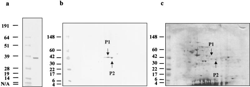
Herpesviruses are associated with several diseases of marine turtles, including lung-eye-trachea disease (LETD) and fibropapillomatosis. Two approaches were used to identify immunodominant antigens of LETV, the LETD-associated herpesvirus. The first approach targeted glycoprotein B, which is known to be immunogenic and neutralizing in other species. The second strategy identified LETV proteins recognized on Western blots by antibodies in immune green turtle plasma. A 38-kDa protein was resolved by two-dimensional gel electrophoresis, sequenced, and identified as a scaffolding protein encoded by the overlapping open reading frames of UL26 and UL26.5. Glycoprotein B and the scaffolding protein were cloned and expressed in Escherichia coli. The expressed proteins were recognized on Western blots by antibodies in immune green turtle plasma. Phylogenetic studies based on UL26, DNA polymerase, and glycoprotein B revealed that LETV clusters with the alphaherpesviruses.


Figures

FIG. 1.
FIG. 1.
Identification of herpesvirus 38-kDa protein by 2D gel electrophoresis and Western blot analysis. The 38-kDa protein was recognized in an LETV-infected cell lysate by plasma from an LETV-immunized green turtle assayed by Western blotting (a). The same plasma sample recognized two protein spots (P1 and P2, indicated by arrows) when the LETV-infected cell lysate was resolved by 2D gel electrophoresis and then assayed by Western blotting (b). The LETV-infected cell lysate was resolved by 2D gel electrophoresis and then Coomassie stained to identify proteins corresponding to the two spots recognized in the Western blot (c). The corresponding proteins, P1 and P2, are indicated by arrows. The values on the left are molecular sizes in kilodaltons. N/A, not available.
FIG. 2.
FIG. 2.
Schematic illustrating the process of obtaining sequence information of gB and of mapping sequences of P1 and P2 to the overlapping UL26 and UL26.5 portion of the genome. (a) Diagram representing a portion of a typical alphaherpesvirus genome. (b) Clones from the LETV partial genomic library containing the corresponding portion of the genome (indicated by hatched boxes). Primers (shown as arrows with numbers) were designed on the basis of the sequence of these clones to PCR amplify the entire open reading frame of the LETV gB gene, as well as additional sequences for UL26.5 and UL26. (c) Diagram representing the two hypothetical independently transcribed messages of the overlapping LETV UL26 and UL26.5 genes (indicated by arrows). (d) The two expected protein products, protease and scaffolding protein, of the putative polyproteins encoded by LETV UL26 (shown along with the anticipated sizes). The two hypothetical cleavage sites are indicated by arrowheads. The protease portion possesses a histidine tag, and the scaffolding protein has a histidine-rich region, as shown. (e) Protein sequences of the 38-kDa protein resolved by 2D electrophoresis (indicated by hatched boxes) aligned with the scaffolding or assembly portion of the overlapping region of UL26 and UL26.5. aa, amino acids.
FIG. 3.
FIG. 3.
Western blot analysis of expressed UL26 (a) and gB (b). In the odd-numbered lanes, lysates of induced E. coli were used as a source of antigen, and in the even-numbered lanes, lysates of the uninduced E. coli control were used as a source of antigen. Molecular weight markers were in lanes MW. Lanes 1 and 2 were incubated with anti-His monoclonal antibody to identify expression of cloned herpesvirus proteins. The LETV scaffolding protein (arrowhead) and protease were recognized by antihistidine antibody. Lanes 3 and 4 were incubated with 1-month post-LETV immunization plasma (a). Lanes 5 and 6 were incubated with plasma from a wild LETV antibody-positive green turtle shown previously to recognize the 38-kDa protein in LETV-infected cell lysates. The expressed LETV glycoprotein (arrowhead) was recognized by antihistidine antibody (b). Lanes 3 and 4 were incubated with 3-month post-LETV immunization plasma. Lanes 5 and 6 were incubated with plasma from an adult LETV antibody-positive green turtle. The values on the left are molecular sizes in kilodaltons.

Similar articles

Cited by

References

    1. Caselli, E., P. G. Balboni, C. Incorvaia, R. Argnani, F. Parmeggiani, E. Cassai, and R. Manservigi. 2001. Local and systemic inoculation of DNA or protein gB1s-based vaccines induce a protective immunity against rabbit ocular HSV-1 infection. Vaccine 19:1225-1231. - PubMed
    1. Coberley, S. S., L. H. Herbst, D. R. Brown, L. M. Ehrhart, D. A. Bagley, S. A. Schaf, R. H. Moretti, E. R. Jacobson, and P. A. Klein. 2001. Detection of antibodies to a disease-associated herpesvirus of the green turtle, Chelonia mydas. J. Clin. Microbiol. 39:3572-3577. - PMC - PubMed
    1. Coberley, S. S., L. H. Herbst, L. M. Ehrhart, D. A. Bagley, S. Hirama, E. R. Jacobson, and P. A. Klein. 2001. Survey of Florida green turtles for exposure to a disease-associated herpesvirus. Dis. Aquat. Org. 47(3):157-167. - PubMed
    1. Corchero, J. L., E. C. Mar, T. J. Spira, P. E. Pellet, and N. Inque. 2001. Comparison of serological assays for detection of antibodies against human herpesvirus 8. Clin. Diagn. Lab. Immunol. 8(5):913-921. - PMC - PubMed
    1. Eggers, M., K. Radsak, G. Enders, and M. Reschke. 2001. Use of recombinant glycoprotein antigens gB and gH for diagnosis of primary human cytomegalovirus infection during pregnancy. J. Med. Virol. 63:135-142. - PubMed

Publication types

MeSH terms

Substances

Associated data

LinkOut - more resources