. 2002 Oct;22(20):6959-70.
doi: 10.1128/MCB.22.20.6959-6970.2002.

The adapter protein ZIP binds Grb14 and regulates its inhibitory action on insulin signaling by recruiting protein kinase Czeta

Affiliations

The adapter protein ZIP binds Grb14 and regulates its inhibitory action on insulin signaling by recruiting protein kinase Czeta

Bertrand Cariou et al. Mol Cell Biol. 2002 Oct.

Abstract

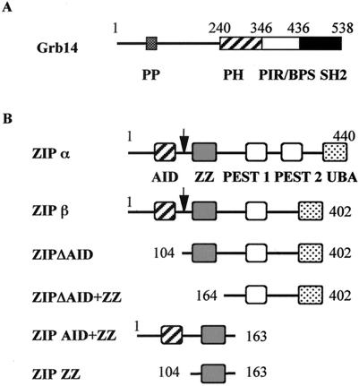
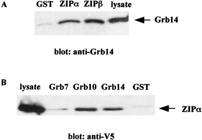
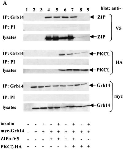
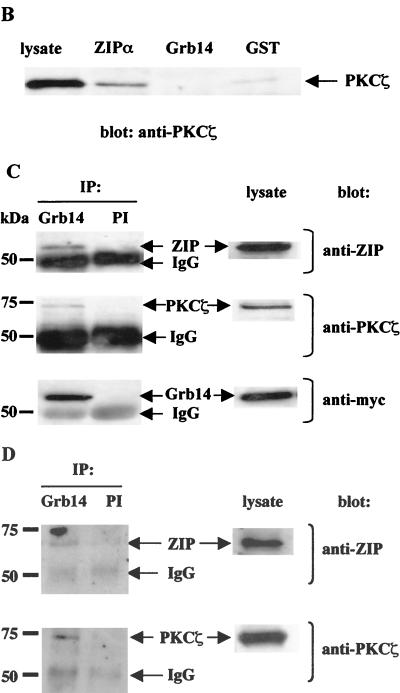
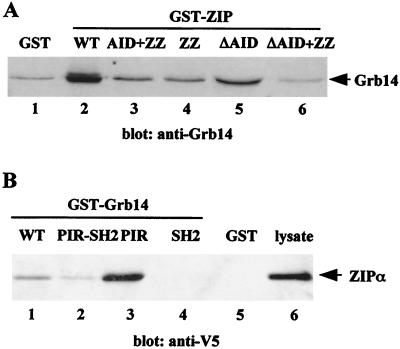
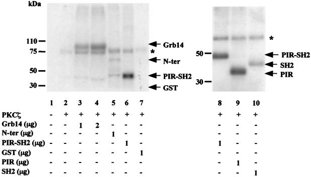
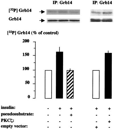
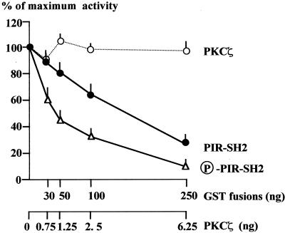
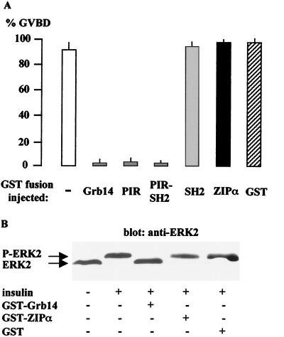
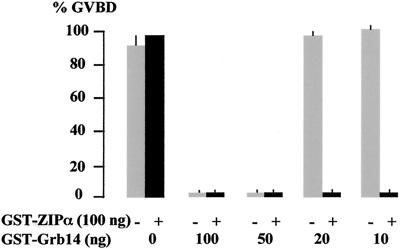
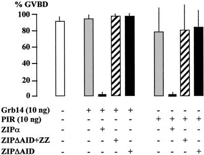
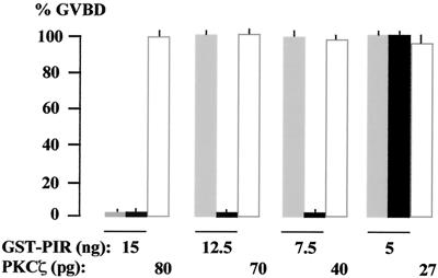
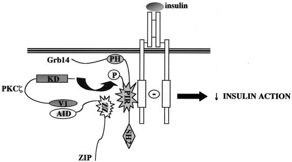
Grb14 is a member of the Grb7 family of adapters and acts as a negative regulator of insulin-mediated signaling. Here we found that the protein kinase Czeta (PKCzeta) interacting protein, ZIP, interacted with Grb14. Coimmunoprecipitation experiments demonstrated that ZIP bound to both Grb14 and PKCzeta, thereby acting as a link in the assembly of a PKCzeta-ZIP-Grb14 heterotrimeric complex. Mapping studies indicated that ZIP interacted through its ZZ zinc finger domain with the phosphorylated insulin receptor interacting region (PIR) of Grb14. PKCzeta phosphorylated Grb14 under in vitro conditions and in CHO-IR cells as demonstrated by in vivo labeling experiments. Furthermore, Grb14 phosphorylation was increased under insulin stimulation, suggesting that the PKCzeta-ZIP-Grb14 complex is involved in insulin signaling. The PIR of Grb14, which also interacts with the catalytic domain of the insulin receptor (IR) and inhibits its activity, was preferentially phosphorylated by PKCzeta. Interestingly, the phosphorylation of Grb14 by PKCzeta increased its inhibitory effect on IR tyrosine kinase activity in vitro. The role of ZIP and Grb14 in insulin signaling was further investigated in vivo in Xenopus laevis oocytes. In this model, ZIP potentiated the inhibitory action of Grb14 on insulin-induced oocyte maturation. Importantly, this effect required the recruitment of PKCzeta and the phosphorylation of Grb14, providing in vivo evidences for a regulation of Grb14-inhibitory action by ZIP and PKCzeta. Together, these results suggest that Grb14, ZIP, and PKCzeta participate in a new feedback pathway of insulin signaling.


Figures

FIG. 1.
FIG. 1.
Schematic representation of Grb14 and ZIP proteins. (A) Grb14. Shown are the proline-rich motif (PP); the PH domain; the PIR (28), also called BPS, between the PH and SH2 domain (22); and the SH2 domain. (B) ZIP and various ZIP constructs. The arrow indicates the length of the ZIP protein isolated in the two-hybrid screen. Shown are the AID (47), ZZ, PEST sequences (PEST2 is lacking in the ZIPβ isoform), and ubiquitin-associated (UBA) domain (70).
FIG. 2.
FIG. 2.
Interaction of Grb14 with ZIP in vitro. (A) ZIP isoforms expressed as GST-fusion proteins or GST control were immobilized on glutathione-Sepharose beads and incubated with lysates of CHO-IR-Grb14 cells. Bound proteins were immunodetected with anti-Grb14 polyclonal antibodies. (B) Grb7 family members expressed as immobilized GST-fusion proteins were incubated with lysates of COS cells transiently transfected with V5 tagged ZIPα. Bound proteins were detected by immunodetection with anti-V5 monoclonal antibodies. These blots are representative of 3 different experiments.
FIG. 3.
FIG. 3.
ZIP forms a ternary complex with Grb14 and PKCζ in vivo. (A) CHO-IR cells were transiently transfected as indicated with 1 μg of either pECE myc-Grb14, pcDNA6 V5-ZIPα, or pcDNA3 HA-PKCζ. pcDNA6 empty plasmid was used to normalize total DNA amount to 3 μg. At 48 h after transfection, cells were stimulated or not with 10−7 M insulin. Cell extracts were immunoprecipitated with polyclonal anti-Grb14 antibodies. Control immunoprecipitations using rabbit preimmune serum were simultaneously performed (PI). The immunoprecipitates were separated by SDS-PAGE and immunoblotted with monoclonal anti-HA, anti-V5, or anti-myc antibodies. Lysates of the same cells were separated by SDS-PAGE and immunoblotted with the same antibodies to confirm equal expressions levels of proteins. (B) Grb14 does not interact with PKCζ in vitro. ZIPα and Grb14 expressed as GST fusion proteins were incubated with human recombinant PKCζ. Bound PKCζ was detected by Western blotting using anti-PKCζ antibodies. (C) Lysates from COS cells transiently transfected with myc-Grb14 were immunoprecipitated with polyclonal anti-Grb14 antibodies. Control immunoprecipitation using rabbit preimmune serum was simultaneously performed. The immunoprecipitates were separated by SDS-PAGE and immunoblotted as indicated with monoclonal anti-ZIP antibodies, anti-myc antibodies, or polyclonal anti-PKCζ antibodies. Lysates of the same cells were analyzed in parallel. (D) Rat liver extracts were immunoprecipitated with polyclonal anti-Grb14 antibodies cross-linked to protein A-agarose. Control immunoprecipitation using rabbit preimmune serum was simultaneously performed. Immunoprecipitates were analyzed by sequential immunodetection using polyclonal anti-ZIP and anti-PKCζ antibodies. In this figure the blots are representative of two or three independent experiments, and lysates and preimmune controls were analyzed on the same gels as experimental samples.
FIG. 3.
FIG. 3.
ZIP forms a ternary complex with Grb14 and PKCζ in vivo. (A) CHO-IR cells were transiently transfected as indicated with 1 μg of either pECE myc-Grb14, pcDNA6 V5-ZIPα, or pcDNA3 HA-PKCζ. pcDNA6 empty plasmid was used to normalize total DNA amount to 3 μg. At 48 h after transfection, cells were stimulated or not with 10−7 M insulin. Cell extracts were immunoprecipitated with polyclonal anti-Grb14 antibodies. Control immunoprecipitations using rabbit preimmune serum were simultaneously performed (PI). The immunoprecipitates were separated by SDS-PAGE and immunoblotted with monoclonal anti-HA, anti-V5, or anti-myc antibodies. Lysates of the same cells were separated by SDS-PAGE and immunoblotted with the same antibodies to confirm equal expressions levels of proteins. (B) Grb14 does not interact with PKCζ in vitro. ZIPα and Grb14 expressed as GST fusion proteins were incubated with human recombinant PKCζ. Bound PKCζ was detected by Western blotting using anti-PKCζ antibodies. (C) Lysates from COS cells transiently transfected with myc-Grb14 were immunoprecipitated with polyclonal anti-Grb14 antibodies. Control immunoprecipitation using rabbit preimmune serum was simultaneously performed. The immunoprecipitates were separated by SDS-PAGE and immunoblotted as indicated with monoclonal anti-ZIP antibodies, anti-myc antibodies, or polyclonal anti-PKCζ antibodies. Lysates of the same cells were analyzed in parallel. (D) Rat liver extracts were immunoprecipitated with polyclonal anti-Grb14 antibodies cross-linked to protein A-agarose. Control immunoprecipitation using rabbit preimmune serum was simultaneously performed. Immunoprecipitates were analyzed by sequential immunodetection using polyclonal anti-ZIP and anti-PKCζ antibodies. In this figure the blots are representative of two or three independent experiments, and lysates and preimmune controls were analyzed on the same gels as experimental samples.
FIG. 4.
FIG. 4.
Mapping of domains involved in the Grb14-ZIP interaction. (A) ZIPα and various ZIP constructs expressed as GST fusion proteins were incubated with lysates of CHO-IR-Grb14 cells. Bound proteins were immunodetected with anti-Grb14 antibodies. (B) Various Grb14 constructs expressed as GST fusion proteins were incubated with lysates of COS cells transiently transfected with V5-ZIPα. Bound proteins were immunodetected using anti-V5 antibodies. Ponceau red staining of the proteins after transfer confirmed that equivalent amounts of GST fusions were loaded. These blots are representative of three different experiments.
FIG. 5.
FIG. 5.
PKCζ phosphorylates Grb14 in vitro. Phosphorylation assays were performed in vitro using human recombinant PKCζ as described in Material and Methods. GST fusion proteins were used as substrate as indicated. Phosphorylated proteins were subjected to SDS-PAGE and analyzed by autoradiography. The asterisk indicates autophosphorylated PKCζ. These blots are representative of four different experiments.
FIG. 6.
FIG. 6.
Grb14 phosphorylation in intact cells. CHO-IR cells transiently transfected with myc-Grb14 and labeled with [32P]orthophosphate were stimulated (+) or not (−) with insulin as indicated. Cells were either cotransfected with HA-PKCζ or empty vector (right part) or treated 30 min with 20 μM of myristoylated PKCζ (Myr-SIYRRGARRWRKL) pseudosubstrate prior to insulin stimulation (left part). Cell lysates were immunoprecipitated with anti-Grb14 antibodies, separated by SDS-PAGE, transferred to nitrocellulose membrane, and quantified with a PhosphorImager (upper blot). Anti-myc immunoblots were then performed to confirm that equal amounts of Grb14 were immunoprecipitated (lower blot). Results are expressed as a percentage of their respective control, in the absence of insulin (left part) or in the absence of HA-PKCζ (right part), and represent the means + standard errors of the means (error bars) of three (right part) or four (left part) independent experiments.
FIG. 7.
FIG. 7.
The phosphorylation of PIR-SH2 of Grb14 increases its inhibitory effect on IR catalytic activity. Tyrosine kinase activity of WGA-purified IR was measured using a synthetic substrate (poly Glu-Tyr 4:1), in the presence of increasing amounts of GST-PIR-SH2 previously phosphorylated or not by PKCζ, or in the presence of PKCζ alone. Results are expressed as a percentage of the maximal insulin effect measured in the absence of GST fusion proteins, and are the means + standard errors of the means of three to five different experiments.
FIG. 8.
FIG. 8.
Grb14 inhibits insulin-induced X. laevis oocyte maturation. Oocytes were microinjected with 100 ng of the indicated GST fusion proteins 1 h before insulin stimulation and analyzed 20 h later for the appearance of GVBD. (A) Effect of the injection of various GST fusions on the insulin induction of oocyte maturation. Results are expressed as a percentage of GVBD. Data represent the means + standard errors of the means (error bars) of two to three animals, each concerning 20 oocytes. (B) Western blot analysis of oocyte lysates using anti-ERK2 antibodies. The blot is representative of three different experiments.
FIG. 9.
FIG. 9.
ZIP potentiates Grb14 inhibition of insulin-induced oocyte maturation. Oocytes were microinjected with decreasing amounts of GST-Grb14 as indicated, together with (filled bars) or without (grey bars) 100 ng of GST-ZIPα. Oocyte maturation in response to insulin was monitored as in Fig. 8. Data are the means + standard errors of the means (error bars) of three animals, each concerning 20 oocytes.
FIG. 10.
FIG. 10.
Importance of Grb14-ZIP-PKCζ interaction in ZIP modulation of Grb14 action. Oocytes were coinjected with GST-Grb14 or GST-PIR in the presence of 100 ng of various GST-ZIP constructs, as indicated. Oocyte maturation in response to insulin was monitored as in Fig. 8. Data are the means + standard errors of the means (error bars) of three animals, each concerning 20 oocytes.
FIG. 11.
FIG. 11.
PIR phosphorylation increases its inhibition of insulin-induced oocyte maturation. Oocytes were either microinjected with the indicated amounts of GST-PIR, which was (black bars) or was not (grey bars) previously phosphorylated by recombinant PKCζ as described in Materials and Methods, or microinjected with PKCζ alone (white bars). Oocyte maturation in response to insulin was monitored as in Fig. 8. Data are the means + standard errors of the means (error bars) of three animals, each concerning 20 oocytes.
FIG. 12.
FIG. 12.
A proposed model for the role of ZIP as an adapter connecting PKCζ to Grb14. ZIP interacts with the V1 domain of PKCζ through its AID domain, and with the PIR of Grb14 through its ZZ zinc finger domain, allowing the formation of a heterotrimeric complex. Under insulin stimulation, the PIR binds to the activated IR regulatory tyrosine kinase loop. PIR phosphorylation by PKCζ leads to an increased Grb14 inhibitory action on IR catalytic activity and insulin action.

References

    1. Bandyopadhyay, G., M. L. Standaert, U. Kikkawa, Y. Ono, J. Moscat, and R. V. Farese. 1999. Effect of transiently expressed atypical (zeta, lambda), conventional (alpha, beta) and novel (delta, epsilon) protein kinase C isoforms on insulin-stimulated translocation of epitope-tagged GLUT4 glucose transporters in rat adipocytes: specific interchangeable effects of protein kinase C-zeta and C-lambda. Biochem. J. 337:461-470. - PMC - PubMed
    1. Bereziat, V., A. Kasus-Jacobi, D. Perdereau, B. Cariou, J. Girard, and A. F. Burnol. 2002. Inhibition of insulin receptor catalytic activity by the molecular adapter Grb14. J. Biol. Chem. 28:4845-4852. - PubMed
    1. Berra, E., M. T. Diaz-Meco, I. Dominguez, M. M. Municio, L. Sanz, J. Lozano, R. S. Chapkin, and J. Moscat. 1993. Protein kinase C zeta isoform is critical for mitogenic signal transduction. Cell 74:555-563. - PubMed
    1. Bossenmaier, B., L. Mosthaf, H. Mischak, A. Ullrich, and H. U. Haring. 1997. Protein kinase C isoforms beta 1 and beta 2 inhibit the tyrosine kinase activity of the insulin receptor. Diabetologia 40:863-866. - PubMed
    1. Braiman, L., A. Alt, T. Kuroki, M. Ohba, A. Bak, T. Tennenbaum, and S. R. Sampson. 2001. Activation of protein kinase C zeta induces serine phosphorylation of VAMP2 in the GLUT4 compartment and increases glucose transport in skeletal muscle. Mol. Cell. Biol. 21:7852-7861. - PMC - PubMed

Publication types

MeSH terms

LinkOut - more resources