. 2002 Oct;22(20):6971-8.
doi: 10.1128/MCB.22.20.6971-6978.2002.

Homologous recombination resolution defect in werner syndrome

Affiliations

Homologous recombination resolution defect in werner syndrome

Yannick Saintigny et al. Mol Cell Biol. 2002 Oct.

Abstract

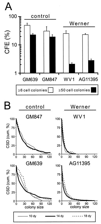
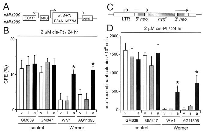
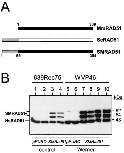
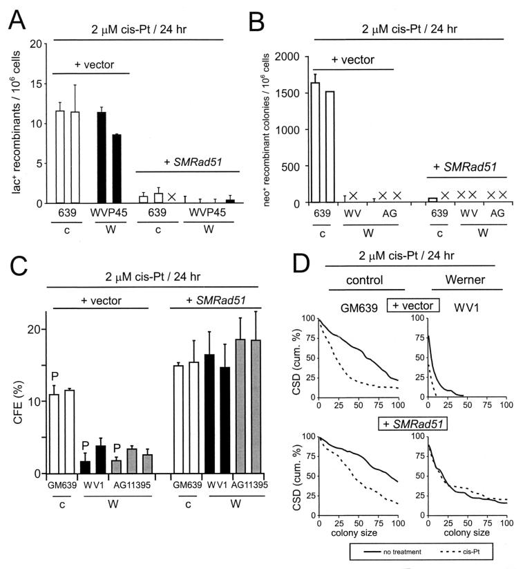
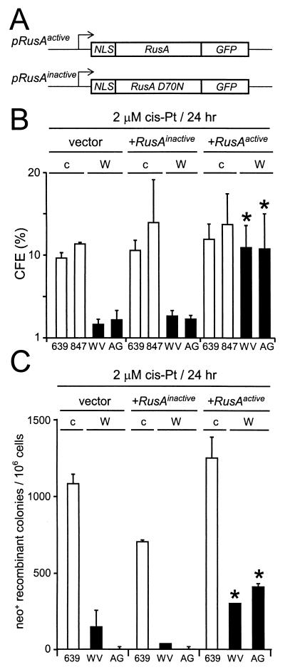
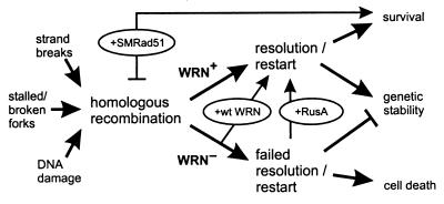
Werner syndrome (WRN) is an uncommon autosomal recessive disease whose phenotype includes features of premature aging, genetic instability, and an elevated risk of cancer. We used three different experimental strategies to show that WRN cellular phenotypes of limited cell division potential, DNA damage hypersensitivity, and defective homologous recombination (HR) are interrelated. WRN cell survival and the generation of viable mitotic recombinant progeny could be rescued by expressing wild-type WRN protein or by expressing the bacterial resolvase protein RusA. The dependence of WRN cellular phenotypes on RAD51-dependent HR pathways was demonstrated by using a dominant-negative RAD51 protein to suppress mitotic recombination in WRN and control cells: the suppression of RAD51-dependent recombination led to significantly improved survival of WRN cells following DNA damage. These results define a physiological role for the WRN RecQ helicase protein in RAD51-dependent HR and identify a mechanistic link between defective recombination resolution and limited cell division potential, DNA damage hypersensitivity, and genetic instability in human somatic cells.


Figures

FIG. 1.
FIG. 1.
Intrinsic growth defect of SV40-transformed WRN cell lines in the absence of DNA damage. (A) CFE of SV40-transformed WRN and control fibroblast cell lines as determined by the ability of single cells to form colonies of ≥6 cells or ≥50 cells after 10 days of growth. The error bars indicate standard deviations for two independent determinations. (B) CSDs for the same four cell lines as determined by the ability of single cells to form colonies of greater than or equal to n cells after 10 to 18 days of growth. The CSD results are plotted as a cumulative distribution (43).
FIG. 2.
FIG. 2.
WRN protein rescues WRN cell survival and recombination after DNA damage. (A) Expression vectors encoding active (pMM290; wild-type [wt] WRN) or missense mutant (pMM289; E84A K577M) WRN protein lacking exonuclease and helicase activities. puror, puromycin resistance gene; EGFP, enhanced green fluorescence protein gene. (B) CFEs of WRN and control cell lines after transfection with control (pPURO) (v), WRN missense mutant (pMM289) (i), or active (pMM290) (a) expression vectors and cis-Pt treatment (2 μM; 24 h). (C) Structure of the direct-repeat recombination reporter plasmid pNeoA (25). The arrows indicate the extent of the neomycin phosphotransferase genes (neo); the solid boxes indicate the positions of inactivating linker insertions, the shaded boxes indicate the overlap region of 592 bp between linker insertion sites, and the hatched box indicates a hygromycin resistance gene (hygr). LTR, long terminal repeat. (D) Frequency of cis-Pt-induced neo+-recombinant colonies per 106 viable cells after transfection as described for panel B. ∗, statistically significant difference in survival or recombinant frequency. The error bars indicate standard deviations for two to five replicate experiments.
FIG. 3.
FIG. 3.
Dominant-negative mammalian RAD51 protein and expression in stable cell lines. (A) Mus musculus (Mm), S. cerevisiae (Sc), and chimeric Mm-Sc (SM) RAD51 proteins (the numbers indicate amino acid residues) (22). Filled and open boxes represent the M. musculus and S. cerevisiae RAD51 open reading frames, respectively. Shaded segments represent the 55 N-terminal amino acid residues of yeast RAD51 that have been fused in frame to mouse RAD51 to generate the chimeric SMRAD51 protein. (B) Western blot analysis of expression of endogenous (HsRAD51) or stably expressed SMRAD51 proteins in representative control (639Rec75) or WRN (WVP46) SV40-transformed fibroblast-derived sublines (35). Lanes 1, 2, 5, and 6 were clonally derived from control plasmid (pPURO) transfections. The two SMRAD51 protein bands arise from alternative initiation or stable degradation (22). Multiple independent sublines were generated for use in subsequent experiments.
FIG. 4.
FIG. 4.
Recombination and cell survival after SMRAD51 protein expression. (A) Frequency of cis-Pt-induced lac+ recombinant cells per 106 viable cells generated by control (+ vector) or SMRAD51-expressing (+ SMRAD51) control (c) and WRN (W) sublines. ×, no cells observed. (B) Frequency of cis-Pt-induced neo+-recombinant colonies per 106 viable cells generated by control or SMRAD51-expressing sublines. ×, no or too few colonies to display using the scale shown (range, 0 to 40 colonies). (C) CFE of WRN and control sublines expressing control plasmid alone or SMRAD51 protein after cis-Pt treatment. The bars represent parental cell lines (P) or clonally derived sublines. The recovery of survival in WRN cells expressing SMRAD51 protein after cis-Pt treatment was highly statistically significant (P < 2 × 10⁻⁵ for all four comparisons; SMRAD51 protein-expressing cells versus the parental cell line or SMRAD51 protein-expressing cells versus the clonally derived subline within both WV1 and AG11395). (D) CSDs of WRN and control sublines expressing vector or SMRAD51 protein after cis-Pt treatment. The recovery of survival of WRN cells expressing SMRAD51 protein after cis-Pt treatment was again highly statistically significant (P < 5 × 10⁻⁷). cum., cumulative. The error bars in all cases indicate standard deviations for two to five replicate experiments.
FIG. 5.
FIG. 5.
Expression of a bacterial resolvase protein improves WRN cell survival and recombination. (A) Vectors for expression of active or catalytically inactive (D70N substitution) forms of the bacterial resolvase protein RusA (NLS, nuclear localization signal; GFP, green fluorescent protein) (11). (B) Survival of control (c) or WRN (W) SV40-transformed fibroblast cell lines transfected with control (vector) plasmid or plasmid expressing inactive (+RusAinactive) or active (+RusAactive) RusA protein and treated with cis-Pt. (C) cis-Pt-induced neo+-recombinant colonies per 106 viable cells after transfection of control (c) or WRN (W) cells as described for panel B. ∗, statistically significant difference compared with control or RusAinactive-transfected cells (see the text). The error bars indicate standard deviations for two to five replicate experiments.
FIG. 6.
FIG. 6.
Model of WRN function and origins of WRN cellular phenotypes. DNA damage, replication, or repair can initiate HR (left) (8, 21, 46). WRN promotes HR resolution or replication restart to insure cell viability and genetic stability (WRN+ arrow). In the absence of WRN (WRN), HR resolution and/or replication restart fails, leading to mitotic arrest, cell death, and genetic instability. Experimental tests of this model are shown in ovals: reexpressing WRN protein (+wt WRN) (Fig. 2) improves both cell survival and the recovery of viable mitotic recombinants, as does expression of the bacterial resolvase protein RusA (+RusA) (Fig. 5). The dependence of WRN phenotypes on RAD51 pathway function and products can be revealed by expressing a dominant-negative form of mammalian RAD51 protein (+SMRAD51) (Fig. 3 and 4) that suppresses mitotic recombination in WRN and controls cells while improving WRN cell survival after cis-Pt damage. Anticipated consequences of survival in the absence of HR function are mutagenesis and genetic instability (45, 46).

References

    1. Bennett, S. E., A. Umar, J. Oshima, R. J. Monnat, Jr., and T. A. Kunkel. 1997. Mismatch repair in extracts of Werner syndrome cell lines. Cancer Res. 57:2956-2960. - PubMed
    1. Boddy, M. N., P.-H. L. Gaillard, W. H. McDonald, P. Shanahan, J. R. I. Yates, and P. Russell. 2001. Mus81-Eme1 are essential components of a Holliday junction resolvase. Cell 107:537-548. - PubMed
    1. Brooks-Wilson, A. R., M. J. Emond, and R. J. Monnat, Jr. 1997. Unexpectedly low loss of heterozygosity in genetically unstable Werner syndrome cell lines. Genes Chromosomes Cancer 18:133-142. - PubMed
    1. Brosh, R. M., Jr., and V. A. Bohr. 2002. Roles of the Werner syndrome protein in pathways required for maintenance of genome stability. Exp. Gerontol. 37:491-506. - PubMed
    1. Chen, X.-B., R. Melchionna, C.-M. Denis, P.-H. L. Gaillard, A. Blasina, I. V. de Weyer, M. N. Boddy, P. Russell, J. Vialard, and C. H. McGowan. 2001. Human Mus81-associated endonuclease cleaves Holliday junctions in vitro. Mol. Cell 8:1117-1127. - PubMed

Publication types

LinkOut - more resources