. 2002 Oct;22(20):7147-57.
doi: 10.1128/MCB.22.20.7147-7157.2002.

Spontaneous access of proteins to buried nucleosomal DNA target sites occurs via a mechanism that is distinct from nucleosome translocation

Affiliations

Spontaneous access of proteins to buried nucleosomal DNA target sites occurs via a mechanism that is distinct from nucleosome translocation

J D Anderson et al. Mol Cell Biol. 2002 Oct.

Abstract

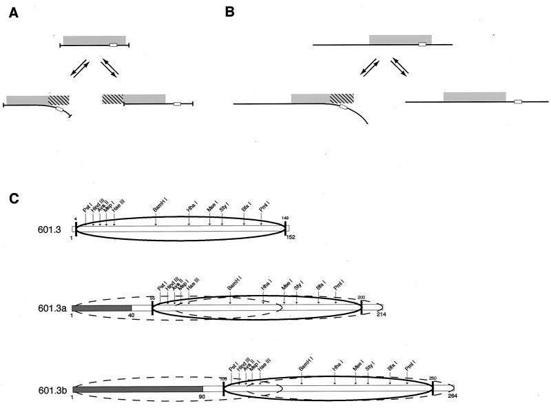
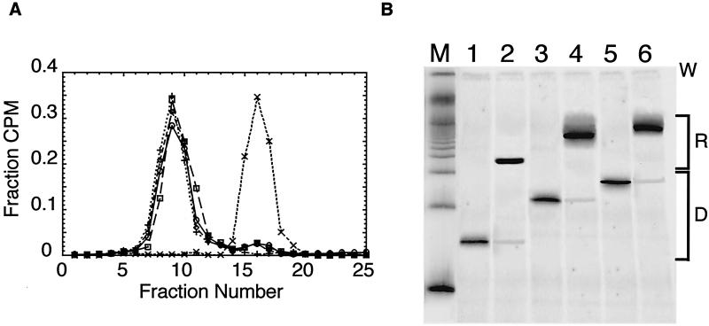
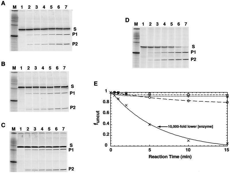
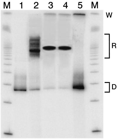
Intrinsic nucleosome dynamics termed "site exposure" provides spontaneous and cooperative access to buried regions of nucleosomal DNA in vitro. Two different mechanisms for site exposure have been proposed, one based on nucleosome translocation, the other on dynamic nucleosome conformational changes in which a stretch of the nucleosomal DNA is transiently released off the histone surface. Here we report on three experiments that distinguish between these mechanisms. One experiment investigates the effects on the accessibilities of restriction enzyme target sites inside nucleosomes when extra DNA (onto which the nucleosome may move at low energetic cost) is appended onto one end. The other two experiments test directly for nucleosome mobility under the conditions used to probe accessibility to restriction enzymes: one on a selected nonnatural nucleosome positioning sequence, the other on the well-studied 5S rRNA gene nucleosome positioning sequence. We find from all three assays that restriction enzymes gain access to sites throughout the entire length of the nucleosomal DNA without contribution from nucleosome translocation. We conclude that site exposure in nucleosomes in vitro occurs via a nucleosome conformational change that leads to transient release of a stretch of DNA from the histone surface, most likely involving progressive uncoiling from an end. Recapture at a distal site along DNA that has partially uncoiled would result in looped structures which are believed to contribute to RNA polymerase elongation and may contribute to spontaneous or ATP-driven nucleosome mobility. Transient open states may facilitate the initial entry of transcription factors and enzymes in vivo.


Figures

FIG. 1.
FIG. 1.
Uncoiling versus translocation mechanisms for DNA site exposure in a nucleosome. (A and B) Schematic illustration of the two mechanisms for nucleosome core particle length DNA (A) or long DNA (B). The left-hand path in each panel illustrates the partial uncoiling mechanism; the right-hand path illustrates the nucleosome translocation mechanism. The histone octamer is shown as a gray rectangle, and the DNA is shown as a line, with a particular target site shown within the nucleosomal DNA (small open rectangle). When the DNA is comparable in length to the 147-bp nucleosome core particle length, then both uncoiling and translocation of the histone octamer lead to net losses of histone-DNA contacts (hatched region of the histone octamer). In this case the two mechanisms are very similar, the major difference being only on which side of the (twofold rotationally symmetric) histone octamer the unsatisfied DNA-binding surface is to be found relative to the side on which a DNA target site is concomitantly made accessible. (C) DNA constructs designed to distinguish between the two mechanisms for site exposure. Sequence 601.3 derives from nucleosome positioning sequence 601.2 (3), after mapping of the nucleosome positioning on 601.2 using exonuclease III; sequences 601.3a and 601.3b extend 601.3 with additional nonnucleosomal 601.2 DNA (open bar) and plasmid DNA (shaded bar) appended. The predominant nucleosome position is illustrated by the heavy ellipse; on sequences 601.3a and 601.3b dashed ellipses indicate limiting alternative positions that could be achieved via nucleosome translocation while maintaining a full set of histone-DNA contacts.
FIG. 2.
FIG. 2.
Purification and characterization of reconstituted nucleosomes. (A) Sucrose gradient purification and reanalysis by sucrose gradient. Nucleosomes are reconstituted by gradual salt dialysis and separated from naked DNA and any nonnucleosomal contaminants on 5 to 30% (wt/vol) sucrose gradients. ○, preparative run of reconstituted 601.3 nucleosomes; □, preparative run of reconstituted 601.3a nucleosomes; ⋄, preparative run of reconstituted 601.3b nucleosomes; +, reanalysis of gradient-purified 601.3 nucleosomes; ×, naked 601.3 DNA. (B) Native gel electrophoresis. W indicates the location of the loading wells, R indicates the mobilities of the reconstituted nucleosomes, and D indicates the mobilities of naked DNA. Lane M, 100 bp DNA marker; lanes 1, 3, and 5, naked DNA for constructs 601.3, 601.3a, and 601.3b, respectively; lanes 2, 4, and 6, the corresponding samples after reconstitution into nucleosomes and purification by sucrose gradient ultracentrifugation. Phosphorimager analysis of the gel reveals contamination of the nucleosomal samples by free DNA to be ≤2%. This small amount of naked DNA does not contribute to the measured Keqconf because it is digested to completion within the first time point, which is omitted from the subsequent analysis.
FIG. 3.
FIG. 3.
Restriction enzyme digestion analyses of site exposure equilibria. Representative data are shown, probing site exposure at a site 100 to 105 bp from the 5′ end of the predominant core particle position. (A to C) Denaturing polyacrylamide gel analysis of the time course of digestion using the enzyme StyI at 1,000 units ml−1. (A) Reconstituted 601.3 nucleosomes; (B) 601.3a nucleosomes; (C) 601.3b nucleosomes. Lanes 1 through 7 are from samples removed at 0, 0.5, 1, 2, 5, 10, and 15 min, respectively, after reaction initiation. In each case, the substrate (S; 152 nucleotides [nt] for construct 601.3, 214 nt for 601.3a and 264 nt for 601.3b) is converted over time into two products (P1, 104 nt, and P2, 44 nt, for 601.3; P1, 154 nt, and P2, 56 nt, for 601.3a; P1, 204 nt, and P2, 56 nt, for 601.3b). The sizes of S, P1, and P2 expected from the DNA sequence are confirmed against the 100-bp DNA markers in lane M. (D) Naked 601.3 DNA digested with StyI at 0.1 units ml−1. (E) Quantitative analysis of the time course of digestion from the data in panels A through D, respectively. The fraction of DNA remaining uncut is plotted versus time. ○, 601.3 nucleosomes; ⋄, 601.3a nucleosomes; □, 601.3b nucleosomes; ×, 601.3 naked DNA. The superimposed lines represent the results of fits to a single exponential plus baseline (see Materials and Methods). Note that 10,000-fold-lower enzyme concentration is used in this naked DNA digestion and that values for Keqconf derive from observed rate constants for digestion scaled by the enzyme concentration. These data show that, were Keqconf to have increased 102- to 104-fold for 601.3a or 601.3b compared to Keqconf for 601.3 at certain sites, this would have been readily apparent.
FIG. 4.
FIG. 4.
Position-dependent equilibrium constants for site exposure (Keqconf) for the three DNA constructs, plotted on a log scale. 601.3, 601.3a, and 601.3b are represented by clear, shaded, and diagonally striped bars, respectively. Values for Keqconf range over 2 orders of magnitude at the sites probed yet are essentially invariant with addition of 55 or 105 bp of DNA beyond the end of the nucleosome.
FIG. 5.
FIG. 5.
Native gel mobility test for nucleosome translocation. Lanes 3 and 4, nucleosomes reconstituted with DNA template 601.3b (264 bp), prior to (lane 3) or after (lane 4) a mock restriction enzyme digestion (30 min, 37°C, HindIII buffer). Lane 2, a (relatively weak) natural nucleosome positioning sequence from the sea urchin 5S rRNA gene (37) (256 bp) yields reconstituted nucleosomes (designated R) at multiple positions on the DNA template, demonstrating that this gel system is capable of resolving such species when they exist. Quantitative studies show that fractions of 0.5% or less of the population having alternative mobilities are readily detected. The fraction of such species present after the mock digestion reaction (lane 4) is undetectably low, and much less than the 10 to 20% of the sample that is actually digested during such reactions when the enzyme is present. Lanes 1 and 5, naked 5S and 601.3b DNA, respectively. D, mobilities of the naked DNA; W, sample loading wells; lanes M, 100-bp ladder size standards.
FIG. 6.
FIG. 6.
Direct test for nucleosome mobility on the 5S nucleosome positioning DNA sequence. (A) Native gel purification of nucleosomes at the preferred and less-favored positions. Lane 1, starting equilibrium distribution of nucleosome positions prepared by salt gradient dialysis; lane 2, gel-purified major positioning isomers (for use as markers); lane 3, gel-purified minor positioning isomers. M, molecular weight markers; D, mobility of naked DNA; R, range of mobilities of reconstituted nucleosomes; W, loading well of the gel. (B) Tests for nucleosome mobility. Nucleosome mobility is manifested by net movement of nucleosomes from the initial disfavored positions (slower mobility) to the dominant (most-favored) major positions (faster mobility). Lanes 1 and 3, purified major isomers, serving as a marker for the electrophoretic mobility of nucleosomes that have moved to the dominant positions; lane 2, positive control showing nucleosome mobility occurring over long times (24 h, TE + 150 mM NaCl, 0°C); lanes 5, 7, 9, and 11, samples after 1 h of incubation at 37°C in NEB restriction enzyme digestion buffers no. 1, 2, 3, and 4, respectively; lanes 4, 6, 8, and 10, same as lanes 5, 7, 9, and 11, except incubated at 0°C. A negligible fraction of nucleosomes move away from the minor positions in 1 h at 37°C in any of the four NEB restriction enzyme buffers, far fewer than are cleaved by restriction enzymes over much shorter times.

References

    1. Anderson, J. D., P. T. Lowary, and J. Widom. 2001. Effects of histone acetylation on the dynamic equilibrium accessibility of nucleosomal DNA target sites. J. Mol. Biol. 307:977-985. - PubMed
    1. Anderson, J. D., and J. Widom. 2001. Poly(dA-dT) promoter elements increase the equilibrium accessibility of nucleosomal DNA target sites. Mol. Cell. Biol. 21:3830-3839. - PMC - PubMed
    1. Anderson, J. D., and J. Widom. 2000. Sequence- and position-dependence of the equilibrium accessibility of nucleosomal DNA target sites. J. Mol. Biol. 296:979-987. - PubMed
    1. Astumian, R. D., and M. Bier. 1994. Fluctuation driven ratchets: molecular motors. Phys. Rev. Lett. 72:1766-1769. - PubMed
    1. Bednar, J., V. M. Studitsky, S. A. Grigoryev, G. Felsenfeld, and C. L. Woodcock. 1999. The nature of the nucleosomal barrier to transcription: direct observation of paused intermediates by electron cryomicroscopy. Mol. Cell 4:377-386. - PubMed

Publication types

LinkOut - more resources