. 2002 Oct;22(20):7204-16.
doi: 10.1128/MCB.22.20.7204-7216.2002.

Pax3-FKHR knock-in mice show developmental aberrations but do not develop tumors

Affiliations

Pax3-FKHR knock-in mice show developmental aberrations but do not develop tumors

Irina Lagutina et al. Mol Cell Biol. 2002 Oct.

Abstract

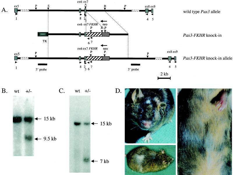
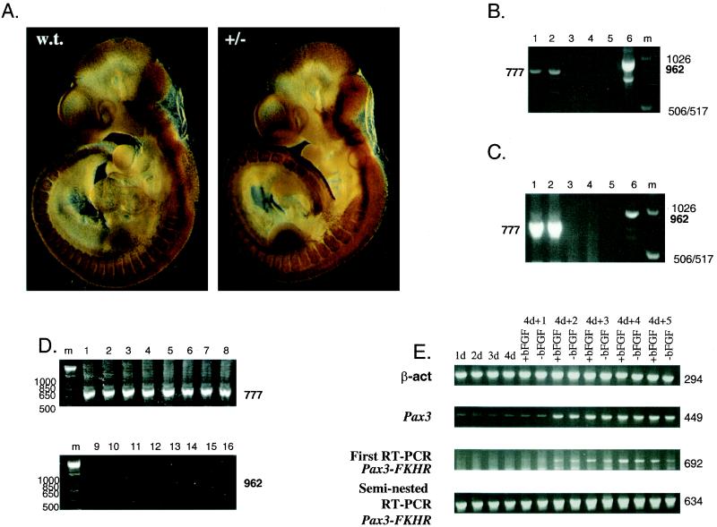
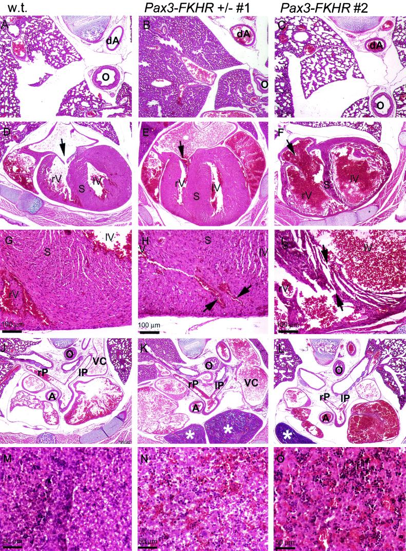
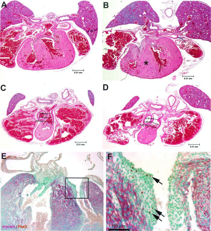
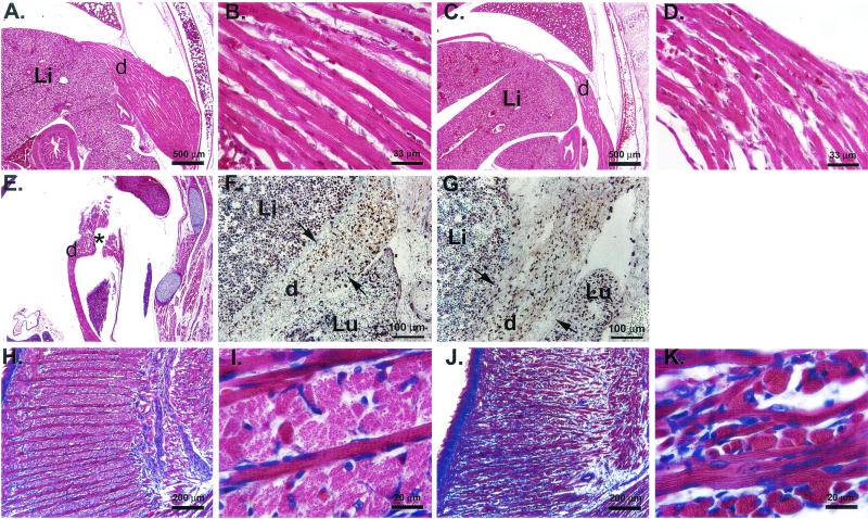
Alveolar rhabdomyosarcoma is a pediatric disease specified by the recurrent chromosome translocations t(2;13) and t(1;13). These translocations result in the formation of the PAX3-FKHR and PAX7-FKHR fusion genes, which are thought to play a causal role in the genesis of this disease. Although PAX3-FKHR exhibits transforming activity in immortalized fibroblast cell lines, a direct role of this fusion protein in tumorigenesis in vivo has not been shown. We determined whether expression of Pax3-FKHR in the mouse germ line would render these animals prone to the development of rhabdomyosarcomas. By targeting FKHR cDNA sequences into the Pax3 locus of embryonic stem cells, we used these cells to generate mice carrying a Pax3-FKHR knock-in allele. Despite low expression of the knock-in allele, heterozygous offspring of Pax3-FKHR chimeric mice showed developmental abnormalities. These included intraventricular septum defects, tricuspid valve insufficiency, and diaphragm defects, which caused congestive heart failure leading to perinatal death. In addition, Pax3-FKHR heterozygous offspring displayed malformations of some but not all hypaxial muscles. However, neither newborn heterozygous pups nor their chimeric parents showed any signs of malignancy. We conclude that the Pax3-FKHR allele causes lethal developmental defects in knock-in mice but might be insufficient to cause muscle tumors.


Figures

FIG.1.
FIG.1.
(A) Generation of the Pax3-FKHR knock-in allele by homologous recombination in ES cells. The top bar represents a physical map of the mouse Pax3 gene encompassing the area around exons 6 and 7 (gray boxes). The middle bar shows the targeting construct in which exon 7 of Pax3 is fused to exon 2 of FKHR (coarsely hatched box) and is followed by a Neor gene (finely hatched box) in the opposite transcriptional orientation. A TK selectable marker (black box) is present at the 5′ end. The bottom line represents the targeted Pax3-FKHR allele. Underneath the targeted allele, the positions of the external 5′ and 3′ probes for Southern blot hybridization are indicated. Horizontal arrowheads indicate the positions of primers, used for genotyping and RT-PCR (Table 1). P, PstI; S, SstI; A, ApaI; B, BamHI; X, XhoI. (B) Southern blot with the 5′ probe, showing wild-type (wt) and targeted Pax3-FKHR (+/−) alleles in a wild-type embryo and a Pax3-FKHR heterozygous littermate, respectively. (C) Southern blot with the 3′ probe showing wild-type (wt) and targeted Pax3-FKHR (+/−) alleles. (D) Middle (upper left) and highly chimeric (lower left and right) Pax3-FKHR chimeric mice, showing white spots on the head, belly, and back.
FIG.2.
FIG.2.
Pax3-FKHR expression in embryos during early gestation and midgestation and in differentiating embryoid bodies. (A) Whole-mount immunostaining of E10.5 wild-type and Pax3-FKHR littermate embryos, using an affinity-purified Pax3 antibody. (B) RT-PCR of heart RNA from E14.5 embryos; even lanes, wild-type RNA; odd lanes, mutant RNA; lanes 1 and 2, first-round RT-PCR for Pax3; lanes 3 and 4, RT-PCR for Pax3-FKHR; lanes 5 and 6, seminested PCR for Pax3-FKHR. (C) RT-PCR of whole RNA from E10.5 embryos. Lanes: 1 and 2, first-round RT-PCR for Pax3 with RNA of wild-type and Pax3-FKHR+/− embryos; 3 and 4, first-round RT-PCR for Pax3-FKHR with RNA of wild-type and Pax3-FKHR+/− embryos; 5 and 6, seminested RT-PCR for Pax3-FKHR with RNA of wild-type and Pax3-FKHR+/− embryos; m, 1-kb marker. (D) RT-PCR of RNA from dissected body parts of E10.5 embryos. Lanes: 1 to 8 first round of RT-PCR for Pax3 with RNA of wild-type (lanes 1 to 4) and Pax3-FKHR+/− (lanes 5 to 8) embryos; 9 to 16 seminested RT-PCR for Pax3-FKHR with RNA of wild-type (lanes 9 to 12) and Pax3-FKHR+/− (lanes 13 to 16) embryos; 1, 5, 9, and 13, neural tubes; 2, 6, 10, and 14 somites and limb buds; 3, 7, 11, and 15, pharyngeal arches; 4, 8, 12, 16, remaining parts of embryos; m, 1-kb marker. Primers: for Pax3, 1 and 4; for first-round Pax3-FKHR RT-PCR, 1 and 7; for seminested Pax3-FKHR PCR, 1 and 6 (Fig. 1A; Table 1). (E) RT-PCR of differentiating embryoid bodies. Lanes: 1d to 4d, RT-PCR of embryoid bodies, grown in suspension for 1 to 4 days; 4d+1 to 4d+5, RT-PCR of embryoid bodies differentiating for 1 to 5 days after growth in suspension, in the presence (+bFGF) or absence (−bFGF) of bFGF; top gel, β-actin gene; second gel from top, Pax3; third gel from top, Pax3-FKHR first-round PCR; bottom gel, Pax3-FKHR second-round PCR. Primers (Fig. 1; Table 1): β-actin gene, 9 and 10; Pax3, 4 and 8; first Pax3-FKHR PCR, 7 and 8; seminested Pax3-FKHR PCR, 6 and 8.
FIG.3.
FIG.3.
Hematoxylin-eosin staining of transverse sections of the thorax region of a P1 wild-type mouse (A, D, G, J, and M) and two Pax3-FKHR heterozygous mice (one dead at birth, [B, E, H, K, and N] and one alive [C, F, I, L, and O]). (A to C) Lungs. Lungs of a wild-type newborn (A), of a dead Pax3-FKHR newborn with noninflated lungs (B) and of a live Pax3-FKHR newborn with inflated lungs (C). (D to F) Heart. Interventricular septa and trabeculated ventricle walls of two mutant hearts appear aberrant (thicker [E] or thinner [F]) compared to those of a wild-type heart (D); the mutant heart (F) also has an abnormally large opening of the tricuspid valve (arrow). (G to I) Interventricular septum. VSD are evident in the hearts of the Pax3-FKHR knock-in mutants (arrows in panels H and I). (J to L) Rostral sections of the heart. The right and left pulmonary arteries are both open and attached to the correct lung; the thymus in mutants has an abnormal, more caudal location (asterisks in panels K and L); and the caval vein is overextended, indicating heart failure (compare VC in panels J and K). (M to O) Liver. The livers of Pax3-FKHR knock-in heterozygous embryos are engorged with blood (N and O), suggestive of heart failure. Abbreviations: rV, right ventricle; lV, left ventricle; S, septum; rP, right pulmonary trunk; lP, left pulmonary trunk; dA, decending aorta; O, esophagus.
FIG. 4.
FIG. 4.
Anatomy of interventricular septum and outflow tract of the hearts of E16.5 wild-type and Pax3-FKHR heterozygous embryos and Pax3 expression. (A to D) Hematoxylin and eosin staining of transverse sections of wild-type (A and C) and mutant (B and D) hearts. The muscular component of the interventricular septum of the heterozygous mutant is already abnormally thickened (asterisk in panel B); the outflow tract of the heart of the Pax3-FKHR mouse is appropriately divided, but the lumen of the pulmonary trunk portion of the outflow tract is dilated (compare panels C and D). (E) Pax3 plus Pax3-FKHR expression in the heart of an E16.5 Pax3-FKHR embryo. (F) Higher magnification of the boxed region of panel E. Green, nuclear staining with methylene green; purple, staining with α-myosin heavy chain; brown, staining with anti-Pax3.
FIG. 5.
FIG. 5.
Muscles of the diaphragm and tongue of Pax3-FKHR heterozygous pups are abnormal around the time of birth but not at E13.5. (A to D) Hematoxylin and eosin staining of similar transverse sections of P1 wild-type (A and B) and Pax3-FKHR knock-in (C and D) diaphragms showing a much reduced musculature of the mutant diaphragm. (E) Sagittal section of a P1 Pax3-FKHR diaphragm showing gaps (asterisk). (F and G) BrdU incorporation and Pax3 and Pax3-FKHR expression in diaphragms of E13.5 wild-type (F) and Pax3-FKHR knock-in (G) pups. Green, nuclear staining with methylene green; purple, staining with anti-BrdU; brown, staining with anti-Pax3. (H to K) Hematoxylin and eosin staining of similar sagittal sections of the tongue of P1 wild-type (H and I) and Pax3-FKHR mutant (J and K) pups. Abbreviations: d, diaphragm; Li, liver; Lu, lung.

References

    1. Anderson, J., T. Gordon, A. McManus, T. Mapp, S. Gould, A. Kelsey, H. McDowell, R. Pinkerton, J. Shipley, and K. Pritchard-Jones. 2001. Detection of the PAX3-FKHR fusion gene in paediatric rhabdomyosarcoma: a reproducible predictor of outcome? Br. J. Cancer 85:831-835. - PMC - PubMed
    1. Anderson, J., A. Ramsay, S. Gould, and K. Pritchard-Jones. 2001. PAX3-FKHR induces morphological change and enhances cellular proliferation and invasion in rhabdomyosarcoma. Am. J. Pathol. 159:1089-1096. - PMC - PubMed
    1. Anderson, M. J., G. D. Shelton, W. K. Cavenee, and K. C. Arden. 2001. Embryonic expression of the tumor-associated PAX3-FKHR fusion protein interferes with the developmental functions of Pax3. Proc. Natl. Acad. Sci. USA 98:1589-1594. - PMC - PubMed
    1. Barr, F. G., N. Galili, J. Holick, J. A. Biegel, G. Rovera, and B. S. Emanuel. 1993. Rearrangement of the PAX3 paired box gene in the paediatric solid tumour alveolar rhabdomyosarcoma. Nat. Genet. 3:113-117. - PubMed
    1. Bennicelli, J. L., S. Advani, B. W. Schafer, and F. G. Barr. 1999. PAX3 and PAX7 exhibit conserved cis-acting transcription repression domains and utilize a common gain of function mechanism in alveolar rhabdomyosarcoma. Oncogene 18:4348-4356. - PubMed

Publication types

MeSH terms