. 2002 Oct;22(20):7279-90.
doi: 10.1128/MCB.22.20.7279-7290.2002.

Endosomal signaling of epidermal growth factor receptor stimulates signal transduction pathways leading to cell survival

Affiliations

Endosomal signaling of epidermal growth factor receptor stimulates signal transduction pathways leading to cell survival

Yi Wang et al. Mol Cell Biol. 2002 Oct.

Abstract

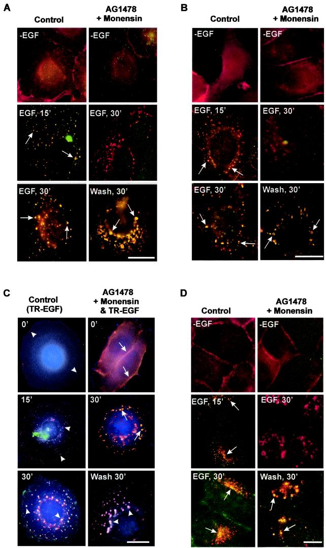
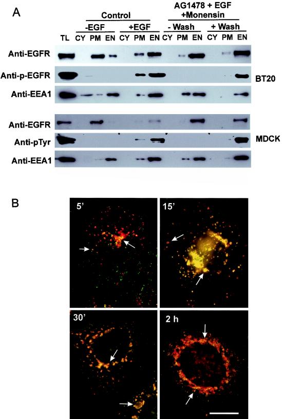
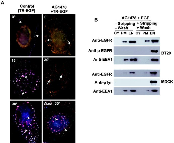
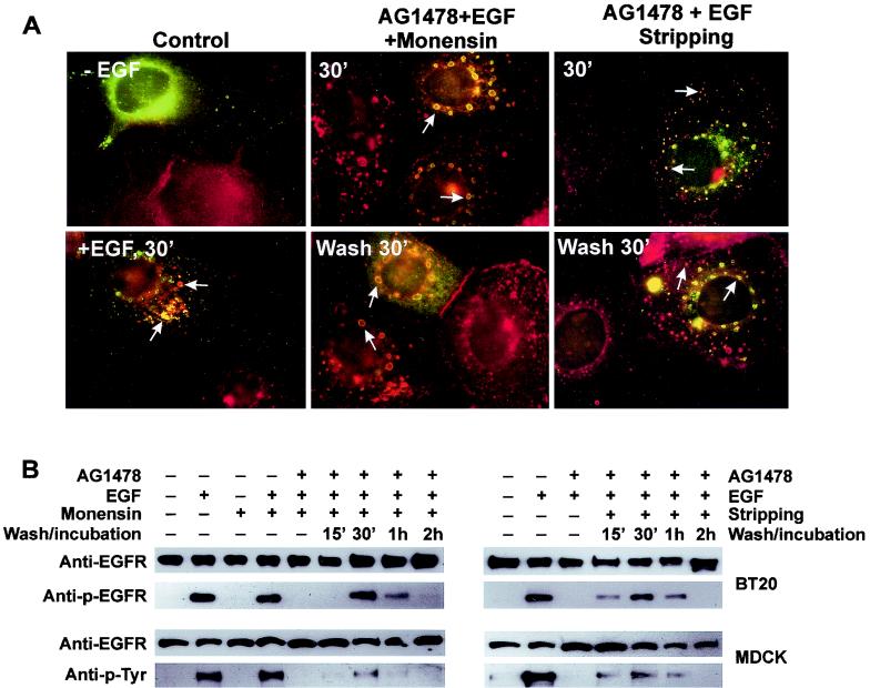
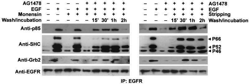
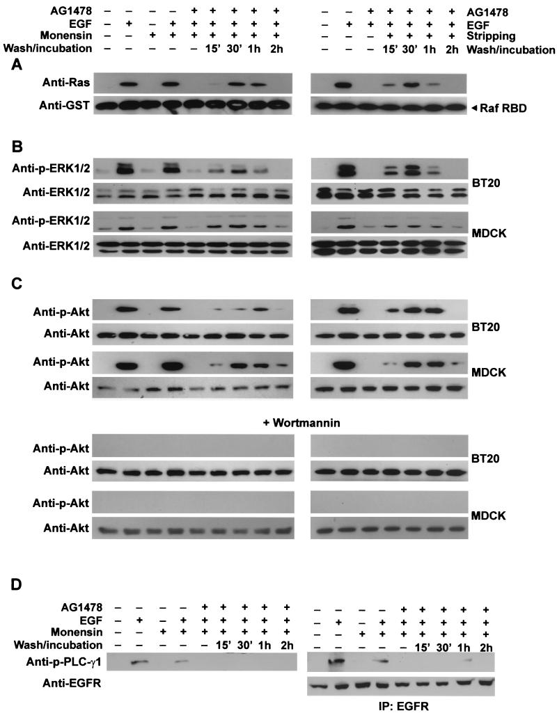
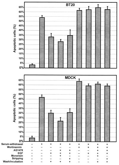
In spite of intensified efforts to understand cell signaling from endosomes, there is no direct evidence demonstrating that endosomal signaling is sufficient to activate signal transduction pathways and no evidence to demonstrate that endosomal signaling is able to produce a biological outcome. The lack of breakthrough is due in part to the lack of means to generate endosomal signals without plasma membrane signaling. In this paper, we report the establishment of a system to specifically activate epidermal growth factor (EGF) receptor (EGFR) when it endocytoses into endosomes. We treated cells with EGF in the presence of AG-1478, a specific EGFR tyrosine kinase inhibitor, and monensin, which blocks the recycling of EGFR. This treatment led to the internalization of nonactivated EGF-EGFR complexes into endosomes. The endosome-associated EGFR was then activated by removing AG-1478 and monensin. During this procedure we did not observe any surface EGFR phosphorylation. We also achieved specific activation of endosome-associated EGFR without using monensin. By using this system, we provided original evidence demonstrating that (i) the endosome can serve as a nucleation site for the formation of signaling complexes, (ii) endosomal EGFR signaling is sufficient to activate the major signaling pathways leading to cell proliferation and survival, and (iii) endosomal EGFR signaling is sufficient to suppress apoptosis induced by serum withdrawal.


Figures

FIG.1.
FIG.1.
Analysis by immunofluorescence of selective activation of EGFR after its endocytosis into endosomes. (A and B) BT20 cells were treated with AG-1478 for 15 min and then stimulated with EGF in the presence of monensin at 37°C for 30 min. Some of the cells were then washed three times with PBS and incubated with serum-free medium for 30 min. BT20 cells that were serum starved or treated only with EGF for 15 and 30 min at 37°C were used as controls; EGFR (red) and pTyr (green) (A) or p-EGFR (green) (B) localization was determined by indirect immunofluorescence. Arrows, colocalization (yellow) of EGFR and pTyr (A) or EGFR and p-EGFR (B). (C) BT20 cells were treated with AG-1478 for 15 min and then stimulated with TR-EGF in the presence of monensin at 4°C for 30 min followed by incubation at 37°C for the indicated times. Some of the cells were then washed with PBS three times and incubated with serum-free medium for the indicated times. BT20 cells that were treated only with TR-EGF at 4°C for 30 min and then at 37°C for indicated times were used as controls; TR-EGF (red), EGFR (green), and pTyr (blue) localization was determined by triple indirect immunofluorescence as described in Materials and Methods. Arrows, colocalization (yellow) of TR-EGF and EGFR; arrowheads, colocalization (light purple) of TR-EGF, EGFR, and pTyr. (D) MDCK cells were treated with AG-1478 for 15 min and then stimulated with EGF in the presence of monensin at 37°C for 30 min. Some of the cells were then washed with PBS three times and incubated with serum-free medium for 30 min. MDCK cells that were serum starved or treated only with EGF for 15 and 30 min at 37°C were used as controls; EGFR (red) and pTyr (green) localization was determined by indirect immunofluorescence. Arrows, colocalization (yellow) of EGFR and pTyr. Bars, 20 μm.
FIG. 2.
FIG. 2.
Further analysis of selective activation of endosome-associated EGFR by subcellular fractionation and by the time course of immunofluorescence. (A) Subcelllular fractionation. BT20 and MDCK cells were treated with AG-1478, EGF, and monensin as described in Materials and Methods. The cells were then subcellularly fractionated into PM, EN, and CY fractions. The subcellular fractions were subjected to immunoblotting with anti-EGFR, anti-pTyr, and anti-EEA1 antibodies. TL, total lysate. (B) Time course by immunofluorescence of the specific activation of endosome-associated EGFR following wash. BT20 cells were treated with AG-1478, EGF, and monensin for 30 min and then washed with PBS for the indicated times. EGFR (red) and pTyr (green) localization was determined by indirect immunofluorescence. Arrows, colocalization (yellow) of EGFR and pTyr. Bar, 20 μm.
FIG. 3.
FIG. 3.
Selective activation of endosome-associated EGFR without monensin. (A) BT20 cells were treated with AG-1478 for 15 min and then stimulated with TR-EGF at 4°C for 30 min, followed by a wash with acidic stripping buffer at 4°C for 1 min. The cells were then washed with PBS three times and incubated with medium at 37°C for the indicated times. BT20 cells were triple fluorescence stained for TR-EGF (red), EGFR (green), and p-EGFR (blue) as described in Materials and Methods. Arrows, colocalization (yellow) of TR-EGF and EGFR; arrowheads, colocalization (light purple) of TR-EGF, EGFR, and p-EGFR. Bar, 20 μm. (B) Subcelllular fractionation. BT20 and MDCK cells were treated with AG-1478 for 15 min and then stimulated with EGF at 37°C for 30 min, followed by a wash with acidic stripping buffer at 4°C for 1 min. The cells were then washed with PBS three times and incubated with medium at 37°C for the indicated times. The cells were then subcellularly fractionated into the PM, EN, and CY fractions. The subcellular fractions were subjected to immunoblotting with anti-EGFR, anti-pTyr, and anti-EEA1 antibodies as described in Materials and Methods. TL, total lysate.
FIG. 4.
FIG. 4.
Trafficking and activity of EGFR following its selective activation in endosomes. (A) Internalization of inactive EGFR into Rab5-positive endosomes and its subsequent activation. BT20 cells were transfected with wild-type Rab5. Cells were either not treated or treated with EGF for 30 min as controls (left). Some cells were treated with AG-1478 for 15 min, followed by addition of EGF and monensin for 30 min without or with washing and incubation (middle). Some cells were treated with AG-1478 and EGF for 30 min without washing or followed by washing with acidic stripping buffer for 1 min and incubation with medium for 30 min. EGFR (red) and Rab5 (green) localization was determined by indirect immunofluorescence. Arrows, colocalization (yellow) of EGFR and Rab5. (B) Immunoblot analysis of the trafficking and inactivation of EGFR following its selective activation in the endosome. BT20 and MDCK cells were treated with AG-1478 and EGF with or without monensin as described for panel A. The cells were washed and then incubated with serum-free medium for the indicated times. Cell lysates were subjected to immunoblot analysis with mouse anti-pTyr, anti-p-EGFR, and rabbit anti-EGFR antibodies.
FIG. 5.
FIG. 5.
Interactions between signaling proteins and endosome-associated EGFR in BT20 cells. (A) BT20 cells were treated with AG-1478 and EGF with or without monensin as described in Materials and Methods. The cells were washed and then incubated with serum-free medium for the indicated times. The cells were lysed and immunoprecipitated (IP) with a rabbit anti-EGFR antibody as described in Materials and Methods. Immunoprecipitates were subjected to immunoblot analysis with mouse anti-EGFR, anti-Grb2, anti-SHC, and anti-p85α antibodies. BT20 cells that were serum starved or treated with EGF, monensin, or EGF plus monensin were used as controls.
FIG. 6.
FIG. 6.
Stimulation of various signal transduction pathways by activation of endosome-associated EGFR. BT20 and MDCK cells were treated with AG-1478 and EGF with or without monensin as described in Materials and Methods. The cells were washed and then incubated with serum-free medium for the indicated times. (A) Activation of Ras. BT20 cell lysates were incubated with GST-RBD conjugated with glutathione beads. The glutathione beads were then subjected to immunoblot analysis with a mouse anti-Ras antibody. GST-Raf RBD was stained by a mouse anti-GST antibody to show equal loading. (B) Activation of ERK. BT20 and MDCK cell lysates were subjected to immunoblot analysis with a rabbit anti-phospho-ERK1/2 (p-ERK1/2) antibody. Total ERK1/2 was used as the control. (C) Activation of Akt and its inhibition by wortmannin. Activation of Akt was determined by an anti-phospho-Akt (p-Akt) antibody. To determine the effects of wortmannin on the activation of Akt, BT20 and MDCK cells were treated with wortmannin (100 nM) for 15 min before the treatment with AG-1478, EGF, and monensin. Cell lysates were subjected to immunoblot analysis with an anti-p-Akt antibody. (D) Activation of PLC-γ1. Activation of PLC-γ1 was determined by immunoblotting of BT20 cell lysates or EGFR immunoprecipitates (IP) with anti-phospho-PLC-γ1 (p-PLC-γ1) antibody.
FIG. 7.
FIG. 7.
Activation of endosome-associated EGFR and the effects on cell survival. BT20 and MDCK cells were incubated in serum-free medium to induce apoptosis. The cells were then treated AG-1478 and EGF (20 ng/ml) with or without monensin followed by washing and incubation as described above. To determine the effects of wortmannin, the cells were pretreated with wortmannin (100 nM). The apoptosis was determined by TUNEL assay.

Similar articles

Cited by

References

    1. Ahn, S., S. Maudsley, L. M. Luttrell, R. J. Lefkowitz, and Y. Daaka. 1999. Src-mediated tyrosine phosphorylation of dynamin is required for beta2-adrenergic receptor internalization and mitogen-activated protein kinase signaling. J. Biol. Chem. 274:1185-1188. - PubMed
    1. Barbieri, M. A., R. L. Roberts, A. Gumusboga, H. Highfield, C. Alvarez-Dominguez, A. Wells, and P. D. Stahl. 2000. Epidermal growth factor and membrane trafficking. EGF receptor activation of endocytosis requires Rab5a. J. Cell Biol. 151:539-550. - PMC - PubMed
    1. Burgering, B. M., and P. J. Coffer. 1995. Protein kinase B (c-Akt) in phosphatidylinositol-3-OH kinase signal transduction. Nature 376:599-602. - PubMed
    1. Burke, P., K. Schooler, and H. S. Wiley. 2001. Regulation of epidermal growth factor receptor signaling by endocytosis and intracellular trafficking. Mol. Biol. Cell 12:1897-1910. - PMC - PubMed
    1. Carpenter, G. 1987. Receptors for epidermal growth factor and other polypeptide mitogens. Annu. Rev. Biochem. 56:881-914. - PubMed

Publication types