. 2002 Oct;22(20):7313-24.
doi: 10.1128/MCB.22.20.7313-7324.2002.

Mouse Af9 is a controller of embryo patterning, like Mll, whose human homologue fuses with Af9 after chromosomal translocation in leukemia

Affiliations

Mouse Af9 is a controller of embryo patterning, like Mll, whose human homologue fuses with Af9 after chromosomal translocation in leukemia

Emma C Collins et al. Mol Cell Biol. 2002 Oct.

Abstract

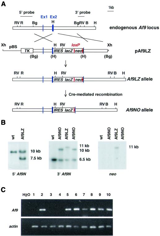
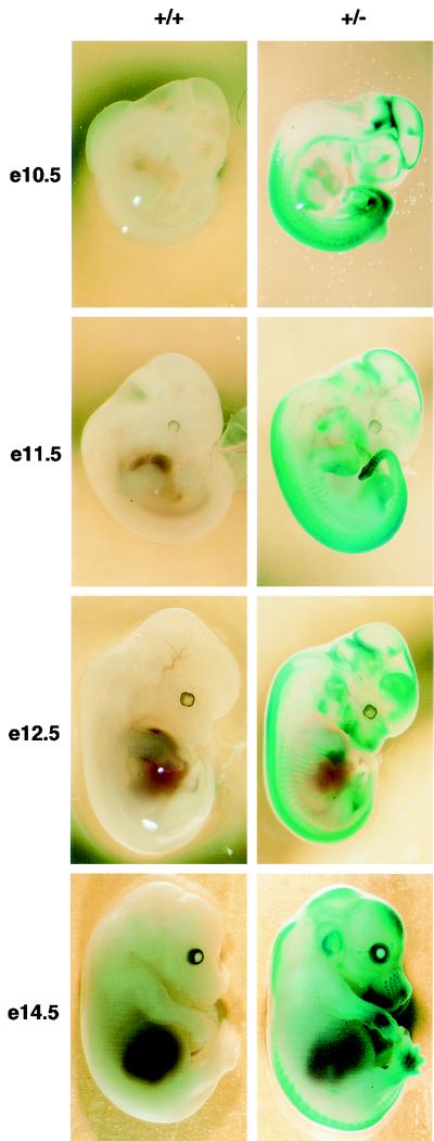
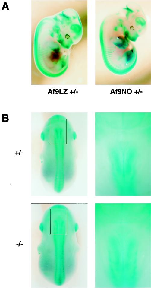
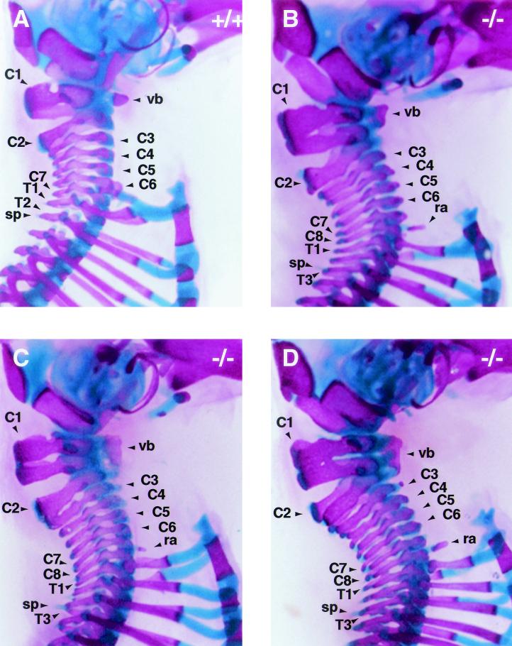
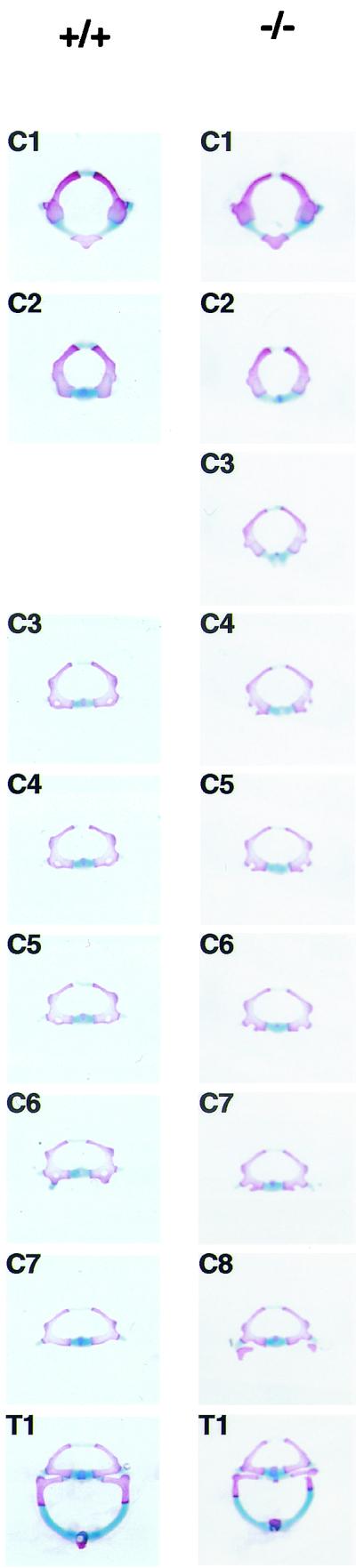
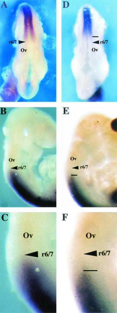
Chromosomal translocation t(9;11)(p22;q23) in acute myeloid leukemia fuses the MLL and AF9 genes. We have inactivated the murine homologue of AF9 to elucidate its normal role. No effect on hematopoiesis was observed in mice with a null mutation of Af9. However, an Af9 null mutation caused perinatal lethality, and homozygous mice exhibited anomalies of the axial skeleton. Both the cervical and thoracic regions were affected by anterior homeotic transformation. Strikingly, mice lacking functional Af9 exhibited a grossly deformed atlas and an extra cervical vertebra. To determine the molecular mediators of this phenotype, analysis of Hox gene expression by in situ hybridization showed that Af9 null embryos have posterior changes in Hoxd4 gene expression. We conclude that the Af9 gene is required for normal embryogenesis in mice by controlling pattern formation, apparently via control of Hox gene regulation. This is analogous to the role of Mll, the murine homolog of human MLL, to which the Af9 gene fuses in acute myeloid leukemias.


Figures

FIG. 1.
FIG. 1.
Targeting strategy for the generation of Af9 null alleles. (A) A partial genomic map of the mouse Af9 gene around the two exons encoding the first 64 amino acids of mouse Af9 (designated exons 1 and 2) is shown in the top line. Targeting vector pAf9LZ (second line) contained a 6.5-kb genomic BglII fragment, with a 5-kb lacZ-neo cassette inserted into a HindIII site in exon 2. The IRES sequence allowed independent translation of the Escherichia coli lacZ gene, which served as an Af9 gene expression marker. The neo sequence conferred G-418 resistance, allowing positive selection of targeted ES cells. This sequence was flanked by loxP sites to allow subsequent excision by transient expression of Cre recombinase (the starting vector was a gift from K. Douglas and A. Smith). Upstream of the targeting fragment, the herpes simplex virus thymidine kinase gene (TK) cassette allowed negative selection of nonhomologous integration events (62). The regions of homology with the endogenous Af9 gene are indicated. The mutant Af9 allele (Af9LZ) resulting from homologous recombination is illustrated in the third line. The Af9NO allele, resulting from transient expression of Cre recombinase, is illustrated in the fourth line. H, HindIII; B, BamHI; Bg, BglII; R, EcoRI; RV, EcoRV; Xh, XhoI. Blue boxes, exons; red arrowheads, LoxP sites. (B) Filter hybridization analysis of ES clones. A 2-kb nonrepetitive EcoRI-BglII fragment 5′ of the targeted region (A) detected a 10-kb EcoRV fragment in the wild-type (wt) genomic DNA, reduced to 7.5 kb in the Af9LZ allele (left). A nonrepetitive 3′ probe consisting of a 1-kb BglII-BamHI fragment (A) detected a 6.5-kb wild-type HindIII band, increased to 11 kb in Af9LZ and then reduced to 10 kb in Af9NO (center). The same 11-kb Af9LZ band hybridized with a probe specific for the inserted neo cassette (right); a single neo insertion was observed. This band was not visible in the Af9NO clones after removal of the neo sequence by expression of Cre recombinase. (C) RT-PCR analysis of RNA from a representative litter of E11.5 Af9NO embryos was used to detect the presence of the Af9 transcript. PCRs were analyzed on agarose gels. PCR carried out with no DNA template (H2O) served as a negative control. A 207-bp band amplified from the Af9 mRNA was generated from all samples, except those from embryos 1, 3, and 4, which were null Af9 mutants. Actin gene-specific RT-PCR, indicating that all cDNA populations analyzed were of comparable quality and concentration, is shown below. Embryo numbers are indicated above the gel. Embryos 1, 3, and 4 were Af9NO−/−, while the remaining embryos were either Af9NO+/− (2, 5, and 7 to 9) or wild type (6 and 10).
FIG. 2.
FIG. 2.
Af9 expression in embryogenesis detected by β-galactosidase staining of Af9LZ mouse embryos. Af9LZ heterozygous mice were mated with C57BL/6 mice, and staged embryos were removed from the uterus, prefixed in 4% PFA, and stained with X-Gal solution overnight. Wild-type embryos (+/+) and heterozygous littermates (+/−) were examined at E10.5, E11.5, E12.5, and E14.5. Activity of the Af9 gene, leading to expression of the β-galactosidase gene in the Af9LZ allele, is visible as blue staining. No staining by endogenous β-galactosidase was observed in the wild-type controls. Areas of precartilage primordium, such as the developing jaw, nose, and skull, as well as the limb buds, developing ribs, and vertebrae, exhibited Af9 expression, particularly at the later stages of development. At E14.5, the external ear and the vibrissae also exhibited strong Af9 expression. Strong staining was also observed in neural tissues, especially the caudal hindbrain, the midbrain-hindbrain junction, and the sympathetic ganglia, as well as in the heart tube, at E10.5.
FIG. 3.
FIG. 3.
Effect of homozygous Af9 mutation on embryonic development. Heterozygous Af9LZ or Af9NO mice were mated, and embryos were removed at E12.5 (A) or E14.5 (B). All embryos were prefixed in 4% PFA and stained with X-Gal solution overnight. (A) Comparison of β-galactosidase staining patterns of Af9LZ and Af9NO heterozygous embryos at E12.5. Similar staining patterns were observed with both lines. (B) Comparison of β-galactosidase staining patterns of heterozygous and homozygous Af9NO embryos. An Af9NO+/− embryo (top) is compared with an Af9NO−/− embryo (bottom; E14.5). A dorsal view of each embryo is shown on the left, with an enlargement of the cervical region on the right. Staining along the axial skeleton extended to a more anterior limit in the homozygous Af9 mutants. In addition, the structures exhibiting Af9 expression appeared to spread over a broader lateral area in this region.
FIG.4.
FIG.4.
Skeletal development in Af9 mutant mice. Skeletons of newborn Af9LZ pups were cleared in 2% KOH and stained for bone with Alizarin red and for cartilage with Alcian blue. A wild-type control mouse (+/+) is shown together with two homozygous Af9 mutant specimens (−/− 1 and 2). Ventral and dorsal views are shown. Among the anomalies observed in the homozygous mutant mice was a lack of the third pair of floating ribs (fr) and malformations of the first and second cervical vertebrae (C1 and C2). Moreover, articulated ribs did not join the sternum (s) pairwise but rather in a staggered, disorganized fashion, resulting in sternum malformations of various levels of severity (see in particular specimen −/− 2). C1, atlas; C2, axis; L1 and L6, first and last lumbar vertebrae, respectively.
FIG. 5.
FIG. 5.
Cervical skeletons of Af9 mutant mice. The cervical regions of skeletons of newborn Af9LZ mice, stained with Alizarin red and Alcian blue, were examined after removal of the forelimbs and shoulder blades. (A) Wild-type (+/+) control mouse; (B to D) homozygous Af9 mutant specimens. The atlases (C1) of Af9 knockout mice were severely deformed; each had the appearance of two vertebrae partially fused, with a single vertebral body (vb) extending along both substructures. Moreover, most Af9 mutants exhibited a complete, supplementary cervical vertebra, bringing the total number of cervical vertebrae to eight, as opposed to the normal seven (B and D). Occasionally, the third vertebra (C3) was partially fused to C2, but with a vertebral body of its own (C). In addition, Af9 knockout mice generally exhibited small protruding rib anlagen (ra) on vertebra C8, whereas wild-type controls usually had a pair of cervical ribs on C6. Furthermore, the spinous process (sp) normally found on the second thoracic vertebra was observed on T3 instead. C1 to C8, cervical vertebrae; T1 to T3, first thoracic vertebrae.
FIG. 6.
FIG. 6.
Morphology of individual vertebrae of Af9 mutant mice. Vertebrae from the cervical and thoracic regions of stained skeletons of newborn Af9LZ mice were dissected and examined under a microscope. Samples from a homozygous Af9 knockout animal (−/−) and the corresponding segments from a wild-type control mouse (+/+) are shown. All cervical vertebrae (C1 to C8) and the first thoracic vertebra (T1) are shown. The third vertebra of Af9 mutants exhibited C2 characteristics, while C4 to C8 resembled C3 to C7 in the wild type.
FIG. 7.
FIG. 7.
Hoxd4 gene expression in Af9 null mutant embryos. Hoxd4 expression was analyzed by whole-mount in situ hybridization (66) of wild type (A to C) and Af9−/− (D to F) E9.5 embryos. A Hoxd4 riboprobe was made from a cDNA fragment and labeled with digoxigenin (46). (A and D) Dorsal views; (B, C, E, and F) lateral views. (C and F) Magnification (×2.5) of the specimens from panels B and E. In wild-type embryos, the Hoxd4 staining extended to a boundary localized between rhombomeres r6 and r7. In the Af9−/− embryos, a posterior shift of the anterior expression limit, corresponding to approximately one rhombomere, was observed. Ov, otic vesicle.

References

    1. Ayton, P. M., and M. L. Cleary. 2001. Molecular mechanisms of leukemogenesis mediated by MLL fusion proteins. Oncogene Rev. 20:5695-5707. - PubMed
    1. Breen, T. R., and P. J. Harte. 1993. Trithorax regulates multiple homeotic genes in the bithorax and Antennapedia complexes and exerts different tissue-specific, parasegment-specific and promoter-specific effects on each. Development 117:119-134. - PubMed
    1. Broeker, P. L., H. G. Super, M. J. Thirman, H. Pomykala, Y. Yonebayashi, S. Tanabe, N. Zeleznik-Le, and J. D. Rowley. 1996. Distribution of 11q23 breakpoints within the MLL breakpoint cluster region in de novo acute leukemia and in treatment-related acute myeloid leukemia: correlation with scaffold attachment regions and topoisomerase II consensus binding sites. Blood 87:1912-1922. - PubMed
    1. Buske, C., and R. K. Humphries. 2000. Homeobox genes in leukemogenesis. Int. J. Hematol. 71:301-308. - PubMed
    1. Cairns, B. R., N. L. Henry, and R. D. Kornberg. 1996. TFG3/TAF30/ANC1, a component of the yeast SW1/SNF complex that is similar to the leukemogenic proteins ENL and AF-9. Mol. Cell. Biol. 16:3308-3316. - PMC - PubMed

Publication types

MeSH terms