. 2002 Oct;22(20):7337-50.
doi: 10.1128/MCB.22.20.7337-7350.2002.

Mechanism of e47-Pip interaction on DNA resulting in transcriptional synergy and activation of immunoglobulin germ line sterile transcripts

Affiliations

Mechanism of e47-Pip interaction on DNA resulting in transcriptional synergy and activation of immunoglobulin germ line sterile transcripts

Sujatha Nagulapalli et al. Mol Cell Biol. 2002 Oct.

Abstract

E47 and Pip are proteins crucial for proper B-cell development. E47 and Pip cooperatively bind to adjacent sites in the immunoglobulin kappa chain 3' enhancer and generate a potent transcriptional synergy. We generated protein-DNA computer models to visualize E47 and Pip bound to DNA. These models predict precise interactions between the two proteins. We tested predictions deduced from these models by mutagenesis studies and found evidence for novel direct interactions between the E47 helix-loop-helix domain (Arg 357 or Asp 358) and the Pip N terminus (Leu 24). We also found that precise spatial alignment of the binding sites was necessary for transcriptional synergy and cooperative DNA binding. A Pip dominant negative mutant that cannot synergize with E47 inhibited enhancer activity in plasmacytoma cells and could not activate transcription in pre-B cells. Using electrophoretic mobility shift assays, we found that Pip can bind to the heavy-chain intron enhancer region. In addition, we found that in fibroblasts Pip greatly increased E47 induction of germ line I micro transcripts associated with somatic rearrangement and isotype class switching. However, a Pip dominant negative mutant inhibited germ line I micro transcripts. The importance of these results for late B-cell functions is discussed.


Figures

FIG. 1.
FIG. 1.
Models of E47 and Pip on the Igκ 3′ enhancer binding sites. (A and B) An E47 bHLH homodimer (red and green) derived from the known E47 crystal structure (14) was positioned on the ideal B-form DNA of the wild-type E-box motif within the Igκ 3′ enhancer. The winged HTH DNA binding domain from IRF-1 was used as a substitute for the Pip DNA binding domain. The known IRF-1 crystal structure (15) was used to model residues 23 to 117 of the Pip DNA binding domain (yellow). Views from the side (A) and bottom (B) of the structure are shown. The arrow shows the N terminus of the IRF structure, which correlates to Pip Lys 23. The sequence of the E47 and Pip motif from the Igκ 3′ enhancer is shown below the model. (C and D) The same E47 and IRF-1 (Pip) DNA binding domains were modeled on the Igκ 3′ enhancer binding sites with a single nucleotide inserted between the E47 and Pip sites. Views from the side (C) and bottom (D) of the structure are shown. The positions of the Pip N terminus and the 1-bp insertion between the E47 and Pip sites are indicated by arrows.
FIG. 2.
FIG. 2.
A single-base-pair insertion destroys E47-Pip synergy. (A) NIH 3T3 cells were transfected with reporter plasmids containing either a multimerized (four copies) E47-Pip motif (lanes 1 to 3) or a multimerized motif with a single-base-pair insertion between the E47 and Pip sites (lanes 4 to 6). Reporter plasmids were cotransfected with E47 or Pip alone or with E47 plus Pip. CAT activities show a dramatic loss in transcriptional synergy by E47 and Pip with the single-base-pair insertion reporter. (B) The single-base-pair insertion did not compromise the E47 binding site. The wild-type and 1-bp insertion reporters were cotransfected with a forced dimer E47 expression vector. Each reporter was equally activated, indicating that the single-base-pair insertion did not reduce the ability of E47 alone to bind to DNA. (C) The single-base-pair insertion does not disrupt separate DNA binding by E47 and Pip. EMSA was performed with wild-type or 1-bp insertion probes with either recombinant E47 or GST-Pip 1-182. Identities of proteins and probes are indicated above the lanes. (D) The single-base-pair insertion reduces E47-Pip cooperative DNA binding. EMSA was performed with either the wild-type E47-Pip DNA binding probe (lanes 1 to 3) or the 1-bp insert probe (lanes 4 to 6). Above each lane are indicated the proteins included in each assay and the probe used. Relative binding values determined by phosphorimager quantification are shown below each lane. (E) All insertions between the E47 and Pip sites in the Igκ 3′ enhancer abolish transcriptional synergy between E47 and Pip. NIH 3T3 cells were transfected with reporter plasmids containing either wild-type (WT) E47-Pip sites or insertions of 1, 2, 5, or 10 bp between the sites. CAT activities are shown in the top panel. Above each lane are shown the identities of cotransfected expression plasmids. The bottom panel shows the E47 and Pip sequences in the reporter plasmids (inserted nucleotides are in boldface), and the histogram shows percent activity of each reporter, with the wild-type reporter activity defined as 100%. Error bars represent standard deviations of the means.
FIG. 3.
FIG. 3.
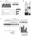
Pip N-terminal sequences are necessary for E47-Pip synergy. (A) Various Pip N-terminal deletion mutants, shown in the bottom panel, were transfected into NIH 3T3 cells along with an E47 expression plasmid and the wild-type (WT) E47-Pip reporter plasmid. CAT activities shown in the top left panel (lanes 1 to 7) indicate that all constructs that lack Pip sequences between residues 20 and 24 show low transcriptional activity. Lanes 8 to 13 show Western blot data for cell extracts of transfected cells probed with anti-Pip antisera, confirming protein expression. The identity of each transfected Pip mutant is shown above each lane. The bottom right panel shows percent activity of each Pip mutant, with wild-type Pip defined as 100%. Error bars represent standard deviations of the means. (B) Deletion of Pip residues 2 to 24 does not affect Pip DNA binding directly but greatly reduces E47-Pip cooperative DNA binding. EMSA with the wild-type E47-Pip DNA probe and various Pip mutant proteins alone or with E47 is shown. The identity of each protein is shown above the lanes. (C) Pip residue Leu 24 is necessary for E47-Pip transcriptional synergy. NIH 3T3 cells were transfected with the E47-Pip reporter plasmid and various Pip mutants either alone (lanes 2 to 7) or with the E47 expression plasmid (lanes 8 to 13). A representative CAT assay is shown in the top panel, and a histogram summarizing the data is shown in the lower right panel. Error bars represent standard deviations of the means. The Pip N-terminal sequence is shown in the bottom left panel. Lanes 14 to 19 show Western blot data for extracts isolated from transfected cells probed with anti-Pip antisera. The identity of each Pip protein is shown above the lanes.
FIG. 4.
FIG. 4.
Identification of E47 residues involved in synergy with Pip. (A) Individual E47 constructs were transfected into NIH 3T3 cells with the E47-Pip-responsive reporter either alone (dark bars) or with the Pip expression plasmid (white bars). CAT activity is normalized to either wild-type E47 alone (dark bars) (defined as 100%) or wild-type E47 plus Pip (white bars) (defined as 100%). The bottom panel shows the E47 helix 1 and loop sequences and the specific amino acids mutated in each construct. Dashes in the bottom panel indicate amino acid identity. (B) Each E47 mutant was prepared in bacteria as a GST fusion protein and used in EMSA with the kappa E47-Pip site as a probe. Identities of proteins added are shown above the lanes.
FIG. 5.
FIG. 5.
Refinement of the E47-Pip (IRF-1)-DNA model. (A and B) The possible effects of DNA bending were modeled by docking the structures of E47 and IRF-1 complexes such that their respective DNA structures were aligned to correspond with that of the Igκ 3′ enhancer. Overlap of the DNA helices can be seen between the two proteins. Shown are side (A) and bottom (B) views of the models. (C) Locations of E47 and Pip residues that disrupt synergy when mutated. The model is viewed from the opposite side as in panels A and B and Fig. 1 to best visualize E47 and Pip residues involved in E47-Pip transcriptional synergy. E47 mutations that affect synergy with Pip are labeled and shown in blue. The position of Pip Leu 24 (IRF-1 Met 8), which is necessary for synergy with E47, is labeled and shown in red.
FIG. 6.
FIG. 6.
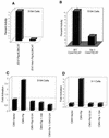
E47-Pip transcriptional synergy in B-cell lines. (A) A single-base-pair insertion between the E47 and Pip sites abolishes endogenous E47-Pip synergy in plasma cells. S194 cells were transfected with either the wild-type E47-Pip-dependent reporter plasmid or the reporter plasmid with a 1-bp insertion between the sites. Activity with the wild-type reporter is defined as 100%. (B) E47-Pip synergy is important for 3′ enhancer activity. Plasmids containing either the entire 3′ enhancer core (WTCoreTKCAT), or with a 1-bp insertion between the E47 and Pip sites (Ins1CoreTKCAT) were transfected into S194 cells. Activity with CoreTKCAT is defined as 100%. (C) Pip mutants that cannot synergize with E47 act as dominant negative mutants in plasmacytoma cells. S194 cells were transfected with the wild-type E47-Pip dependent reporter plasmid and either wild-type Pip expression plasmid or mutant Pip expression plasmids. The identity of each Pip expression plasmid is shown below the lanes. (D) Pip mutants that cannot synergize with E47 fail to activate transcription in pre-B cells. 3-1 pre-B cells were transfected with the wild-type E47-Pip-dependent reporter plasmid and various Pip expression constructs. The identity of each Pip expression plasmid is shown below the lanes.
FIG. 7.
FIG. 7.
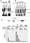
E47-Pip synergy at the Ig heavy-chain locus. (A) The heavy-chain μE4 site is a weak E47-Pip motif. EMSA was performed with either the kappa E47-Pip DNA probe or the μE4 site probe. Proteins added are indicated above the lanes. The arrow points to a weak DNA-Pip complex with the μE4 probe. (B) Pip binds to the heavy-chain intron enhancer region. EMSA was performed with a 220-bp HinfI DNA fragment containing the entire heavy-chain intron enhancer core. Proteins added are indicated above the lanes. The mass in micrograms of Pip 1-182 added is shown at the top. Arrows point to either the Pip 1-182-DNA complex or the E47-Pip-DNA complex. (C) E47-Pip synergy stimulates endogenous sterile Iμ transcripts. NIH 3T3 cells were transfected with plasmids expressing E47, Pip, E47 plus Pip, or E47 plus PipΔ2-24. RNA was isolated from transfected cells and subjected to RT-PCR with primers specific for sterile Iμ or actin transcripts. The top panel shows a map of the heavy-chain locus and diagrams Iμ transcripts. The middle panel shows results of RT-PCR assays using fourfold-increasing amounts of cDNA with either Iμ-specific or actin-specific primers. The data in the middle panel were quantitated by phosphorimager analysis, normalized for transfection efficiency by β-galactosidase activity, and then normalized to the actin control. The histogram in the bottom panel shows the normalized level of Iμ transcripts in each lane. The identities of the constructs used in each transfection are shown above the lanes.

Similar articles

Cited by

References

    1. Bain, G., E. C. R. Maandag, D. J. Izon, D. Amsen, A. M. Kruisbeek, B. C. Weintraub, I. Krop, M. S. Schlissel, A. J. Feeney, M. van Roon, M. van der Valk, H. P. J. te Riele, A. Berns, and C. Murre. 1994. E2A proteins are required for proper B cell development and initiation of immunoglobulin gene rearrangements. Cell 79:885-892. - PubMed
    1. Bain, G., and C. Murre. 1998. The role of E-proteins in B- and T-lymphocyte development. Semin. Immunol. 10:143-153. - PubMed
    1. Bain, G., W. J. Romanow, K. Albers, W. L. Havran, and C. Murre. 1999. Positive and negative regulation of V(D)J recombination by the E2A proteins. J. Exp. Med. 189:289-300. - PMC - PubMed
    1. Benezra, R., R. L. Davis, D. Lockshon, D. L. Turner, and H. Weintraub. 1990. The protein Id: a negative regulator of helix-loop-helix DNA binding proteins. Cell 61:49-59. - PubMed
    1. Betz, A. G., C. Milstein, A. Gonzalel-Fernandez, R. Pannell, T. Larson, and M. S. Neuberger. 1994. Elements regulating somatic hypermutation of an Igκ gene: critical role of the intron enhancer/matrix attachment region. Cell 77:239-248. - PubMed

Publication types

MeSH terms

LinkOut - more resources