. 2002 Oct 1;99(20):12753-8.
doi: 10.1073/pnas.162488899. Epub 2002 Sep 19.

Insig-2, a second endoplasmic reticulum protein that binds SCAP and blocks export of sterol regulatory element-binding proteins

Affiliations

Insig-2, a second endoplasmic reticulum protein that binds SCAP and blocks export of sterol regulatory element-binding proteins

Daisuke Yabe et al. Proc Natl Acad Sci U S A. .

Abstract

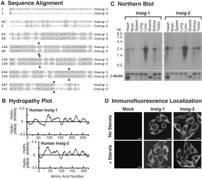
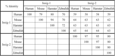
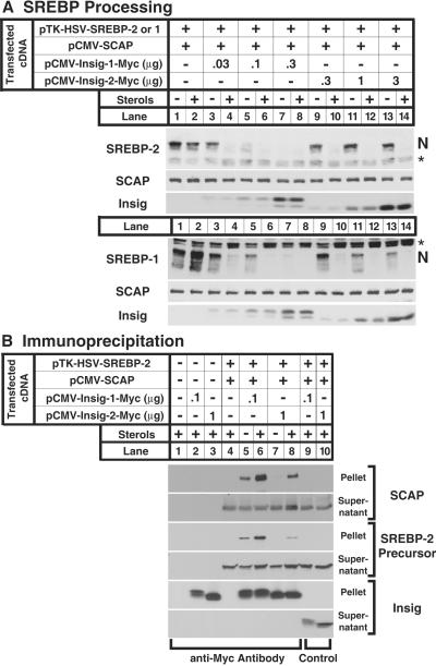
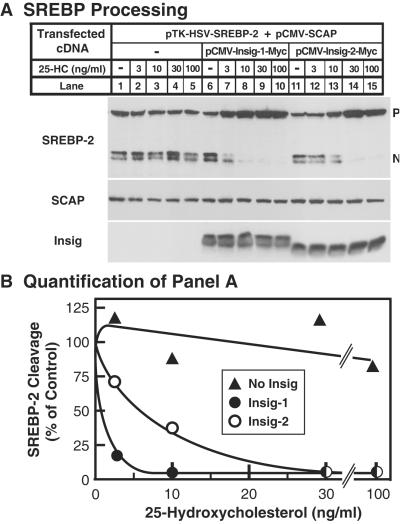
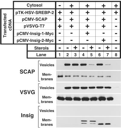
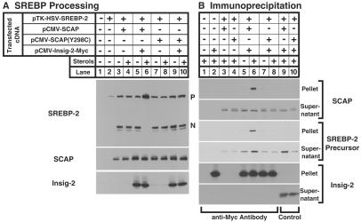
This paper describes insig-2, a second protein of the endoplasmic reticulum that blocks the processing of sterol regulatory element-binding proteins (SREBPs) by binding to SCAP (SREBP cleavage-activating protein) in a sterol-regulated fashion, thus preventing it from escorting SREBPs to the Golgi. By blocking this movement, insig-2, like the previously described insig-1, prevents the proteolytic processing of SREBPs by Golgi enzymes, thereby blocking cholesterol synthesis. The sequences of human insig-1 and -2 are 59% identical. Both proteins are predicted to contain six transmembrane helices. The proteins differ functionally in two respects: (i) production of insig-1, but not insig-2, in cultured mammalian cells requires nuclear SREBPs; and (ii) at high levels of expression, insig-1, but not insig-2, can block SCAP movement in the absence of exogenous sterols. The combined actions of insig-1 and -2 permit feedback regulation of cholesterol synthesis over a wide range of sterol concentrations.


Figures

Figure 1
Figure 1
Characteristics of human insig-1 and -2. (A) Amino acid sequence alignment. Identical residues are shaded. Junctions between exons are indicated by triangles. GenBank accession numbers for human insig-1 and -2 are AY112745 and AF527632, respectively. (B) Hydropathy plot. The residue-specific hydropathy index was calculated over a window of 18 residues (23) by using software from DNASTAR. (C) Northern blot analysis of insig-1 and -2 mRNAs in mouse tissues. Filters containing poly(A)+ RNA (2 μg per lane) from mouse tissues (CLONTECH) were hybridized with 32P-labeled probes against mouse insig-1 and mouse insig-2 (1.5 × 10⁶ cpm/ml for each probe) for 1 h at 68°C as described in Supporting Materials and Methods. Filters were exposed to Kodak X-Omat films with intensifying screens at −80°C for 18 h. The same filters were subsequently hybridized with a 32P-labeled probe against human β-actin (1.5 × 10⁶ cpm/ml) and exposed to film for 3 h at −80°C. (D) Immunofluorescence localization of human insig-1 and -2 in stably transfected CHO cells. On day 0, mock-transfected CHO-7 cells (Mock), CHO/pInsig-1-Myc cells (insig-1), and CHO/pInsig-2-Myc cells (insig-2) were seeded onto coverslips (4 × 10⁴ cells per 37-mm well) in medium B. On day 2, the cells were washed twice with PBS and switched to medium C in the absence (no sterols) or presence of 1 μg/ml of 25-hydroxycholesterol plus 10 μg/ml of cholesterol (+ sterols). On day 3, the cells were fixed, and indirect immunofluorescence was carried out as described (7). (Scale bars, 10 μm.)
Figure 2
Figure 2
Amino acid sequence conservation among insig proteins from various species. Sequences of insig-1 and -2 from human, mouse, hamster, and zebrafish were aligned against each other by using the ALIGN program (www2.igh.cnrs.fr/bin/align-guess.cgi; ref. 24). The percentages of identical amino acids are shown. GenBank accession numbers: AY112745 (human insig-1); AF527630 (mouse insig-1); AF527628 (hamster insig-1); AF527626 (zebrafish insig-1); AF527632 (human insig-2); AF527631 (mouse insig-2); AF527629 (hamster insig-2); and AF527627 (zebrafish insig-2).
Figure 3
Figure 3
Actions of insig-1 and insig-2 in transfected hamster cells. (A) Restoration of sterol-mediated inhibition of SREBP-1 and -2 cleavage in cells overexpressing SCAP. On day 0, SRD-13A cells were set up in medium B at 4 × 10⁵ cells per 60-mm dish. On day 2, the cells were transfected with the indicated plasmids: 2 μg of pTK-HSV-SREBP-2 (Upper) or pTK-HSV-SREBP-1a (Lower), 1 μg of pCMV-SCAP, and the indicated amounts of pCMV-Insig-1-Myc or pCMV-Insig-2-Myc. On day 3, cells were switched to medium C containing 1% (wt/vol) hydroxypropyl-β-cyclodextrin and incubated for 1 h at 37°C. Cells were then washed twice with PBS and switched to medium C in the absence or presence of 1 μg/ml 25-hydroxycholesterol plus 10 μg/ml cholesterol (sterols). After incubation for 4 h, cells were harvested, and total cell extracts were prepared as described (11). Aliquots of extracts (50 μg) were subjected to SDS/PAGE and immunoblot analysis with IgG-1D2 (SREBP-2), IgG-2A4 (SREBP-1), IgG-9D5 (SCAP), and IgG-9E10 (insig) as indicated. N denotes the cleaved nuclear form of SREBP-2 or -1. Asterisk denotes a cross-reactive band. (B) Coimmunoprecipitation of insig-1 and -2 with SCAP and SREBP-2. On day 0, SRD-13A cells were set up in medium B at 8 × 10⁵ cells per 100-mm dish. On day 2, the cells were transfected as in A. On day 3, cells were treated as in A. After incubation for 4 h, cells were harvested for immunoprecipitation with polyclonal anti-Myc IgG (lanes 1–8) or control nonimmune IgG (lanes 9, 10) as described in Materials and Methods. Immunoprecipitated pellets (representing 0.25 dish of cells) and supernatants (0.05 dish of cells) were subjected to SDS/PAGE and immunoblot analysis with IgG-1D2 (SREBP-2), IgG-9D5 (SCAP), and IgG-9E10 (insig).
Figure 4
Figure 4
Insig-1 and -2 increase sensitivity of SREBP processing to inhibition by 25-hydroxycholesterol. (A) On day 0, SRD-13A cells were set up in medium B at 4 × 10⁵ cells per 60-mm dish. On day 2, the cells were transfected with the indicated plasmids: 2 μg of pTK-HSV-SREBP-2, 1 μg of pCMV-SCAP, and either 0.1 μg of pCMV-Insig-1-Myc or 1 μg of pCMV-Insig-2-Myc. On day 3, cells were switched to medium C containing 1% hydroxypropyl-β-cyclodextrin and incubated for 1 h at 37°C. Cells were then washed twice with PBS and switched to medium C plus the indicated concentration of 25-hydroxycholesterol (25-HC). After incubation for 4 h, total cell extracts were prepared. Aliquots of the extracts (50 μg) were subjected to SDS/PAGE and immunoblot analysis with IgG-1D2 (SREBP-2), IgG-9D5 (SCAP), and IgG-9E10 (insig). N and P denote the cleaved nuclear and uncleaved precursor forms of SREBP-2, respectively. (B) The gels in A were scanned and quantified by densitometry. The intensity of the cleaved nuclear form of SREBP-2 in lanes 1, 6, and 11 was arbitrarily set at 100%.
Figure 5
Figure 5
Restoration of sterol-mediated inhibition of SCAP budding from ER membranes in vitro by insig-1 and -2. On day 0, SRD-13A cells were set up in medium B at 8 × 10⁵ cells per 100-mm dish and grown at 37°C. On day 2, the cells were transfected with the indicated plasmids: 2 μg of pTK-HSV-SREBP-2, 1 μg of pCMV-SCAP, 1 μg of pVSVG-T7, and either 0.1 μg of pCMV-Insig-1-Myc or 1 μg of pCMV-Insig-2-Myc. After incubation at 40°C for 12 h, the cells were switched to medium C containing 1% (wt/vol) hydroxypropyl-β-cyclodextrin and incubated for 1 h at 40°C. Cells were then washed twice with PBS and switched to medium C in the absence or presence of 1 μg/ml 25-hydroxycholesterol plus 10 μg/ml cholesterol (sterols) as indicated. After incubation for 4 h at 40°C, cells were harvested. Membranes were incubated in vitro under conditions that permit vesicle budding as described in figure 8 of Nohturfft et al. (11). Aliquots (80 μg protein) of the membranes were incubated in the absence (lane 1) or presence (lanes 2–6) of rat liver cytosol for 15 min at 28°C. Vesicle and membrane fractions were separated by centrifugation, and aliquots containing 100% of the vesicle protein and 15% of the membrane protein were subjected to SDS/PAGE and immunoblot analysis with IgG-9D5 (SCAP), anti-T7⋅Tag antibody (VSVG), and IgG-9E10 (Insig).
Figure 6
Figure 6
Sterol-dependent interaction of insig-2 with wild-type SCAP, but not with sterol-resistant mutant SCAP(Y298C). (A) On day 0, SRD-13A cells were set up in medium B at 4 × 10⁵ cells per 60-mm dish. On day 2, the cells were transfected with the indicated plasmids: 2 μg of pTK-HSV-SREBP-2, 1 μg of either pCMV-SCAP or pCMV-SCAP(Y298C), and 1 μg of pCMV-Insig-2-Myc. On day 3, cells were switched to medium C containing 1% hydroxypropyl-β-cyclodextrin and incubated for 1 h at 37°C. Cells were then washed twice with PBS and switched to medium C in the absence or presence of 0.1 μg/ml 25-hydroxycholesterol plus 10 μg/ml cholesterol (sterols). After incubation for 4 h, cells were harvested. Aliquots of cell extracts (50 μg) were subjected to SDS/PAGE and immunoblot analysis with IgG-1D2 (SREBP-2), IgG-9D5 (SCAP), and IgG-9E10 (insig-2). N and P denote the cleaved nuclear and uncleaved precursor forms of SREBP-2, respectively. (B) On day 0, SRD-13A cells were set up in medium B at 8 × 10⁵ cells per 100-mm dish. On day 2, the cells were transfected as in A. On day 3, cells were treated as in A. After incubation for 4 h, cells were harvested for immunoprecipitation with polyclonal anti-Myc IgG (lanes 1–8) or control nonimmune IgG (lanes 9, 10) as described in Materials and Methods. Immunoprecipitated pellets (representing 0.25 dish of cells) and supernatants (0.05 dish of cells) were subjected to SDS/PAGE and immunoblot analysis with IgG-1D2 (SREBP-2), IgG-9D5 (SCAP), and IgG-9E10 (insig-2).
Figure 7
Figure 7
SREBPs regulate expression of insig-1 mRNA, but not insig-2 mRNA in hamster cells. (A) On day 0, N-BP cell lines were set up at 1 × 10⁶ cells per 100-mm dish in medium B. On day 2, the cells were switched to medium A supplemented with 5% lipoprotein-deficient serum and the indicated amount of ponasterone A. After incubation for 24 h at 37°C, the cells were harvested for preparation of total RNA. Total RNA (10 μg per lane) was subjected to electrophoresis and hybridized for 1 h at 68°C with 32P-labeled probes against hamster insig-1, hamster insig-2, and human β-actin (1.5 × 10⁶ cpm/ml for each probe) as described in Supporting Materials and Methods. Filters were exposed to films with intensifying screens at −80°C for 2 days (insig-1), 7 days (insig-2), or 3 h (β-actin). (B) On day 0, cells were set up in medium B at the following density: 5 × 10⁵ cells per 100-mm dish (CHO-K1, CHO/pS2P, and SRD12B cells) and 8 × 10⁵ cells per 100-mm dish (M19 and SRD13A cells). On day 2, the cells were switched to medium C in the absence or presence of 1 μg/ml of 25-hydroxycholesterol plus 10 μg/ml of cholesterol (sterols). After incubation for 18 h at 37°C, the cells were harvested, and total RNA was analyzed as described in A. Filters were exposed to films at −80°C for 5 days (insig-1 and -2) and 1 h (β-actin).

References

    1. Brown M S, Goldstein J L. Proc Natl Acad Sci USA. 1999;96:11041–11048. - PMC - PubMed
    1. Edwards P A, Tabor D, Kast H R, Venkateswaran A. Biochim Biophys Acta. 2000;1529:103–113. - PubMed
    1. Goldstein J L, Rawson R B, Brown M S. Arch Biochem Biophys. 2002;397:139–148. - PubMed
    1. Horton J D, Goldstein J L, Brown M S. J Clin Invest. 2002;109:1125–1131. - PMC - PubMed
    1. Brown, A. J., Sun, L., Feramisco, J. D., Brown, M. S. & Goldstein, J. L. (2002) Mol. Cell, in press. - PubMed

Publication types

MeSH terms