. 2002 Oct 1;99(20):13067-72.
doi: 10.1073/pnas.202491599. Epub 2002 Sep 19.

Aberrant accumulation of EFEMP1 underlies drusen formation in Malattia Leventinese and age-related macular degeneration

Affiliations

Aberrant accumulation of EFEMP1 underlies drusen formation in Malattia Leventinese and age-related macular degeneration

Lihua Y Marmorstein et al. Proc Natl Acad Sci U S A. .

Abstract

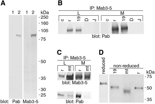
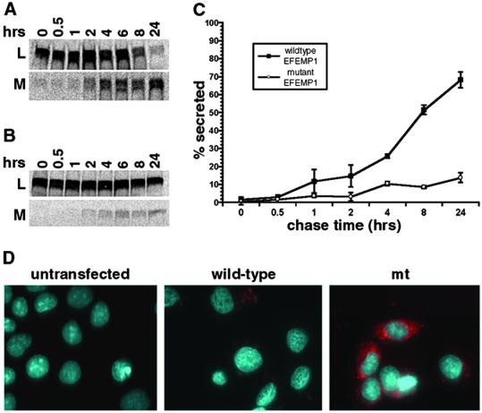
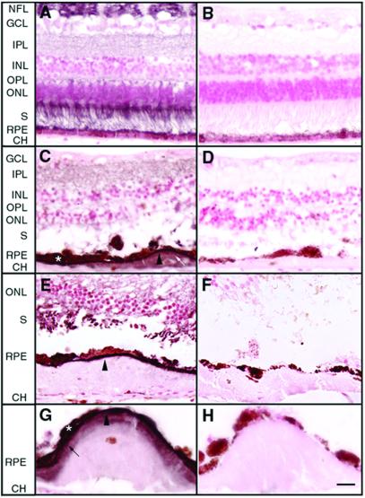
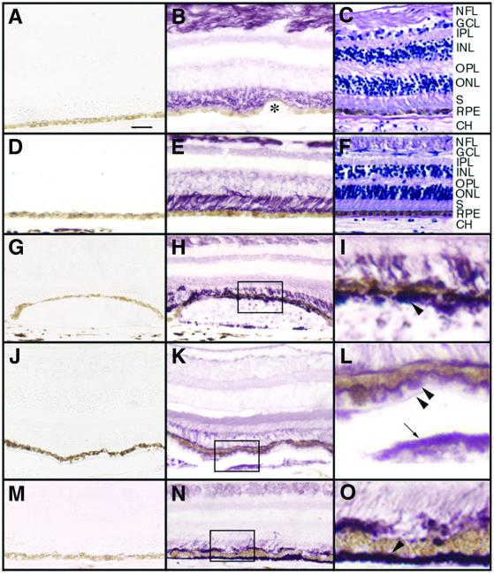
Malattia Leventinese (ML), an inherited macular degenerative disease, is closely reminiscent of age-related macular degeneration (AMD), the most common cause of incurable blindness. Both ML and AMD are characterized by extracellular deposits known as drusen between the retinal pigment epithelium (RPE) and Bruch's membrane. The mechanism underlying drusen formation is unknown. An Arg to Trp mutation in a gene of unknown function, EFEMP1, is responsible for ML, indicating EFEMP1 may be important in drusen formation. Here, we show that wild-type EFEMP1 is a secreted protein whereas mutant EFEMP1 is misfolded, secreted inefficiently, and retained within cells. In normal eyes, EFEMP1 is not present at the site of drusen formation. However, in ML eyes, EFEMP1 accumulates within the RPE cells and between the RPE and drusen, but does not appear to be a major component of drusen. Furthermore, in AMD eyes, EFEMP1 is found to accumulate beneath the RPE immediately overlaying drusen, but not in the region where there is no apparent retinal pathology observed. These data present evidence that misfolding and aberrant accumulation of EFEMP1 may cause drusen formation and cellular degeneration and play an important role in the etiology of both ML and AMD.


Figures

Figure 1
Figure 1
Characterization of EFEMP1 antibodies and wild-type and mutant EFEMP1 protein. (A) Lysates of 293 cells transfected with control vector (lane 1) or EFEMP1-RFP DNA (lane 2) were immunoblotted with Pab or Mab3-5 antibody against EFEMP1. The only band detected in the blots corresponds to the EFEMP1-RFP fusion protein (84 kDa) in the EFEMP1-RFP transfected cell lysates, indicating the high specificity of these antibodies. (B), Cell lysates (L) and media (M) of 293 cells transfected with control vector (lane c) or EFEMP1 cDNA (lane r), or untransfected ARPE-19 (lane 19), D407 (lane D), and RPE-J (lane J) cells were immunoprecipitated with Mab3-5 and blotted with Pab. A band with a relative molecular mass of ≈55 kDa was detected in both the lysates and media of 293 cells transfected with EFEMP1 cDNA and ARPE-19 cells. (C) Lysates (L) and media (M) of 293 cells transfected with EFEMP1 cDNA without (lane r) or with the R345W mutation (lane mt) were immunoprecipitated with Mab3-5 and blotted with Mab3-5 (Upper) or Pab (Lower). Note mutant EFEMP1 comigrates with wild-type EFEMP1. There is noticeably more mutant EFEMP1 in the lysates than the media compared with wild-type EFEMP1. Note Pab recognizes mutant EFEMP1 poorly. (D) Endogenous EFEMP1 in ARPE-19 medium (lane 19) and recombinant wild-type (lane r) and mutant (lane mt) EFEMP1 expressed in 293 transfectant media were analyzed under nonreducing conditions (non-reduced). A control of EFEMP1 under reducing conditions is shown (reduced). Note that mutant EFEMP1 migrates faster than the wild type in nonreducing conditions.
Figure 2
Figure 2
Comparison of wild-type and mutant EFEMP1 secretion. Pulse–chase secretion assays were performed for EFEMP1 in RPE-J cells transfected with the full-length EFEMP1 cDNA without (A) or with the R345W mutation (B). (C) The percentage of secretion in pulse–chase secretion assays was derived from the protein amount in the medium divided by the combining amount in the lysate and medium at each time point. By 24 h of chase, nearly 70% of the wild-type EFEMP1 was in the medium, whereas only 13% of mutant EFEMP1 was secreted. (D) Transfected RPE-J cells stained with monoclonal antibody 3-5 (red). The nuclei were stained with 4′,6-diamidino-2-phenylindole (DAPI; blue).
Figure 3
Figure 3
The distribution of EFEMP1 in normal or ML eyes. Paraffin sections of human donor eyes were probed with Mab3-5 or a control mouse monoclonal antibody RET-PE2 in equivalent dilutions to Mab3-5. The control antibody recognizes a species-specific epitope in rat extracellular matrix metalloproteinase inducer (EMMPRIN). Diaminobenzidine (DAB; brownish color) was used as substrate, and nuclei were counterstained with nuclear fast red. (A and B) Normal eye from a 55-yr-old male (A) was stained with Mab3-5 and (B) the control antibody. Note that EFEMP1 staining is predominantly associated with the nerve fiber layer (NFL) and the photoreceptor inner and outer segments (S). Less intense staining was observed in the outer nuclear layer (ONL) and the inner (IPL) and outer (OPL) plexiform layers. No staining was observed in the RPE, Bruch's membrane, the choroid (CH), or the inner nuclear layer (INL). (CH) Eye from an 86-yr-old female ML patient homozygous for the R345W mutation in EFEMP1 gene. C, E, and G were stained with Mab3-5, and D, F, and H with the control antibody. An intense line of EFEMP1 staining separates the RPE from the drusen beneath (C, E, and G, arrowheads). Some RPE cells overlaying drusen were heavily stained by Mab3-5 (C and G, asterisk). A few drusen were stained by the EFEMP1 antibody (C, white arrow) although the intensity of the staining was always lower than the region between the RPE and drusen. In some drusen, a defined region adjacent to the RPE appeared stained, with the remainder of the deposits apparently devoid of EFEMP1 (G, black arrow). GCL, ganglion cell layer. [Scale bar = 40 μm (AF) and 20 μm (G and H).]
Figure 4
Figure 4
The distribution of EFEMP1 in normal or AMD eyes. Paraffin sections were probed with Mab3-5 (B, E, H, I, K, L, N, and O) or the control mouse monoclonal antibody RET-PE2 (A, D, G, J, and M). Vector VIP (purple color) was used as substrate, and nuclei were not counterstained. Some sections were stained by hematoxylin/eosin (C and F) to illustrate the morphology. (AC) Normal eye from a 91-yr-old female. Again, note that EFEMP1 staining is predominantly associated with the nerve fiber layer (NFL) and the photoreceptor inner and outer segments (S). No staining was observed in the RPE, Bruch's membrane, the choroid (CH), or small hard drusen (B, asterisk). (DF) The peripheral region of the AMD eyes where no pathology was observed. Note the EFEMP1 staining (E) is similar to that in normal eyes (B). (GI) The macular region of an AMD eye. I is higher magnification of the box area in H. Note intense EFEMP1 staining beneath the RPE immediately overlaying a soft druse (H and I, arrowhead). (JL) The macular region of a second AMD eye. L is higher magnification of the box area in K. Again, note the heavy EFEMP1 staining beneath the RPE overlaying soft drusen (K and L, arrowheads). In this eye, drusen were also stained by the EFEMP1 antibody (K and L, arrow), and there is EFEMP1 staining condensed along the apical surface of the RPE, leaving the photoreceptor inner and outer segment staining less intense. (MO) The macular region of a third AMD eye. O is higher magnification of the boxed area in N. Note heavy EFEMP1 staining of basal deposits (N and O, arrowhead). There is also heavier EFEMP1 staining at the apical surface of the RPE and less intense staining in the photoreceptor inner and outer segment region. GCL, ganglion cell layer; IPL, inner plexform layer; INL, inner nuclear layer; OPL, outer plexform layer; ONL, outer nuclear layer. [Scale bar = 50 μm (AH, J, K, M, and N) and 12.5 μm (I, J, and O).]

References

    1. Doyne R W. Trans Ophthalmol Soc UK. 1899;19:71.
    1. Vogt A. In: Untersuchungsmethoden. 3rd Ed. Graefe A, Saemisch T, editors. Berlin: Verlag von Wilhelm Engelman; 1925. pp. 1–118.
    1. Klainguti R. Klin Monatsbl Augenheikd. 1932;89:253–254.
    1. Evans K, Gregory C Y, Wijesuriya S D, Kermani S, Jay M R, Plant C, Bird A C. Arch Ophthalmol. 1997;115:904–910. - PubMed
    1. Collins T. Ophthalmoscope. 1913;11:537–538.

Publication types

MeSH terms