. 2002 Sep 26;21(43):6624-32.
doi: 10.1038/sj.onc.1205910.

Cell type-specific responses of human cells to inhibition of replication licensing

Affiliations

Cell type-specific responses of human cells to inhibition of replication licensing

S Shreeram et al. Oncogene. .

Abstract

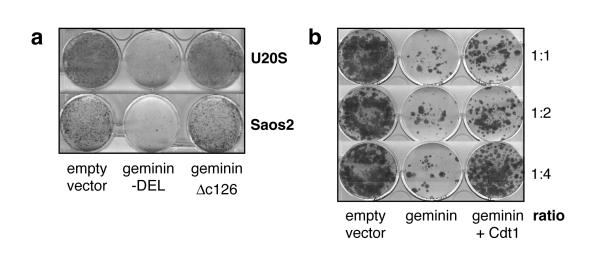
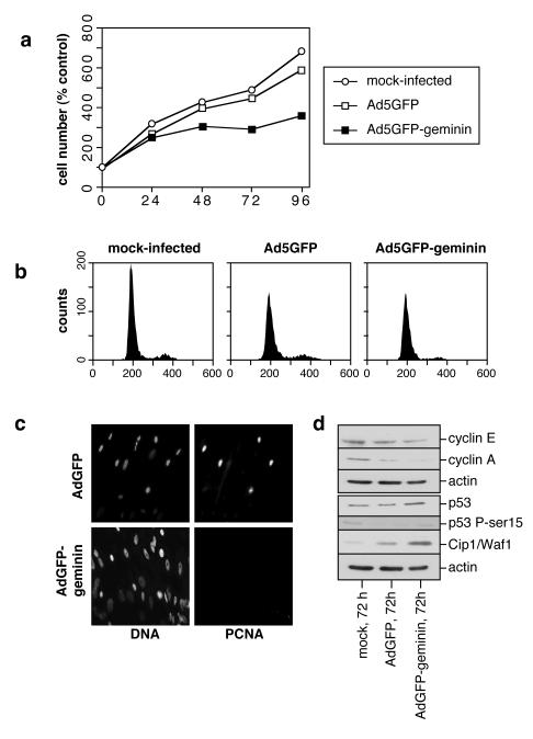
Replication origins are 'licensed' for a single initiation event by loading Mcm2-7 complexes during late mitosis and G1. Licensing is blocked at other cell cycle stages by the activity of cyclin-dependent kinases and a small protein called geminin. Here, we describe the effects of over-expressing a non-degradable form of geminin in various cell lines. Geminin expression reduced the quantity of Mcm2 bound to chromatin and blocked cell proliferation. U2OS (p53+/Rb+) cells showed an early S phase arrest with high cyclin E and undetectable cyclin A levels, consistent with the activation of an intra-S checkpoint. Saos2 (p53-/Rb-) cells showed an accumulation of cells in late S and G2/M with approximately normal levels of cyclin A, consistent with loss of this intra-S phase checkpoint. Geminin also induced apoptosis in both these cell lines. In contrast, IMR90 primary fibroblasts over-expressing geminin arrested in G1 with reduced cyclin E levels and no detectable apoptosis. A 'licensing checkpoint' may therefore act in primary cells to prevent passage into S phase in the absence of sufficient origin licensing. These results suggest that inhibition of the licensing system may cause cancer-specific cell killing and therefore represent a novel anti-cancer target.


Figures

Figure 1
Figure 1
Inhibition of cell proliferation by geminin and rescue by Cdt1. a, U2OS cells were transfected with empty pcDNA3 vector, pcDNA3 containing a stable form of geminin (geminin-DEL) or pcDNA3 containing an inactive form of geminin (gemininΔc126). Cells were grown under selection for 3 weeks and colonies were then stained with Giemsa. b, U2OS cells were transfected with empty pcDNA3, or pcDNA3 expressing geminin-DEL plus or minus pcDNA3 expressing Cdt1 at a ratio of 1:1, 1:2 or 1:4. Cells were grown under selection for 3 weeks and then stained with Giemsa.
Figure 2
Figure 2
Expression of geminin by adenovirus vector. a, U2OS, Saos2 and IMR-90 cells were treated by mock infection or infection with Ad5GFP-geminin. Geminin expression was assessed by immunoblotting cell extracts 72 h and 96 h later. Blots were also probed with actin antibodies as a loading control. b, U2OS, Saos2 and IMR90 cells were infected with either Ad5GFP or Ad5GFP-geminin. After 72 h, total cell extracts were prepared (T) and fractionated into a soluble supernatants (S) or chromatin-containing pellets (P), and then immunoblotted for Mcm2.
Figure 3
Figure 3
Analysis of adenovirus-infected U2OS. U2OS cells were infected with either Ad5GFP or Ad5GFP-geminin or were mock-infected. a, At different times, cell number was measured and expressed as the percentage change over mock-infected controls at time 0 h. Numbers represent the mean of three independent experiments. b, 72 h (top panel) or 96 h (bottom panel) after infection cellular DNA was stained with propidium iodide and analysed by flow cytometry.
Figure 4
Figure 4
PCNA and BrdU staining of adenovirus-infected U2OS. U2OS cells were infected with either Ad5GFP or Ad5GFP-geminin and were incubated for 96 h. a, Cells were fixed and PCNA was detected by indirect immunofluorescence with an anti-PCNA antibody and fluorescein-labelled secondary antibody. DNA was stained with DAPI. b, Cells were pulsed for 30 min with BrdU, fixed and detected with an anti-BrdU antibody and Texas red labelled secondary antibody. DNA was stained with DAPI.
Figure 5
Figure 5
Cell cycle status of adenovirus-infected U2OS. U2OS cells were infected with either Ad5GFP or Ad5GFP-geminin or were mock-infected. a, At 72 or 96 h (72 h for mock-infected cells) cell extracts were prepared and stained with antibodies against cyclin E, cyclin A, p53, p53 phospho-serine 15, Cip1/Waf1 or actin. b, An in situ TUNEL assay was performed 96 h post infection. Total DNA was stained with DAPI.
Figure 6
Figure 6
Cell cycle analysis in U2OS cells infected with increasing MOI of Ad5GFP-geminin. U2OS cells were infected with either Ad5GFP or Ad5GFP-geminin or were mock-infected, at the indicated MOI. 96 h after infection cellular DNA was stained with propidium iodide and analysed by flow cytometry.
Figure 7
Figure 7
Analysis of adenovirus-infected Saos2. Saos2 cells were infected with either Ad5GFP or Ad5GFP-geminin or were mock-infected. a, At different times, cell number was measured and expressed as the percentage change over mock-infected controls at time 0 h. Numbers represent the mean of three independent experiments. b, 96 h after infection cellular DNA was stained with propidium iodide and analysed by flow cytometry. c, At 96 h, infected cells were pulsed for 30 min with BrdU, fixed and detected with an anti-BrdU antibody and Texas red labelled secondary antibody. DNA was stained with DAPI. d, At 72 or 96 h cell extracts were prepared and immunoblotted with antibodies against cyclin E, cyclin A or actin.
Figure 8
Figure 8
Analysis of Ad5GFP-geminin infected primary fibroblasts. IMR90 cells were infected with either Ad5GFP or Ad5GFP-geminin or were mock-infected. a, At different times, cell number was measured and expressed as the percentage change over mock-infected controls at time 0 h. Numbers represent the mean of three independent experiments. b, 72 h after infection cellular DNA was stained with propidium iodide and analysed by flow cytometry. c, At 72 h cells were fixed and PCNA was detected by indirect immunofluorescence with an anti-PCNA antibody and fluorescein-labelled secondary antibody. DNA was stained with DAPI. d, At 72 h cell extracts were prepared and stained with antibodies against cyclin E, cyclin A, p53, p53 phospho-serine 15, Cip1/Waf1 or actin. Note that the p53 phospho-serine 15 blot was produced in parallel with the one in Fig 5a and is shown at the same exposure.

References

    1. Abraham RT. Genes Dev. 2001;15:2177–2196. - PubMed
    1. Arentson E, Faloon P, Seo J, Moon E, Studts JM, Fremont DH, Choi K. Oncogene. 2002;21:1150–1158. - PubMed
    1. Blow JJ, Hodgson B. Trends Cell Biol. 2002;12:72–78. - PMC - PubMed
    1. Blow JJ, Laskey RA. Nature. 1988;332:546–548. - PubMed
    1. Burkhart R, Schulte D, Hu D, Musahl C, Gohring F, Knippers R. Eur. J. Biochem. 1995;228:431–438. - PubMed

Publication types