

Quantitative magnetic resonance imaging of the brain in survivors of very low birth weight
- PMID: 12243993
- PMCID: PMC1763037
- DOI: 10.1136/adc.87.4.279
Quantitative magnetic resonance imaging of the brain in survivors of very low birth weight
Abstract
Background: Children who survive very low birth weight (VLBW) without major disability have a high prevalence of learning difficulty, attention deficit, and dyspraxia.
Aims: To determine whether learning difficulty in children with VLBW is associated with structural brain abnormalities.
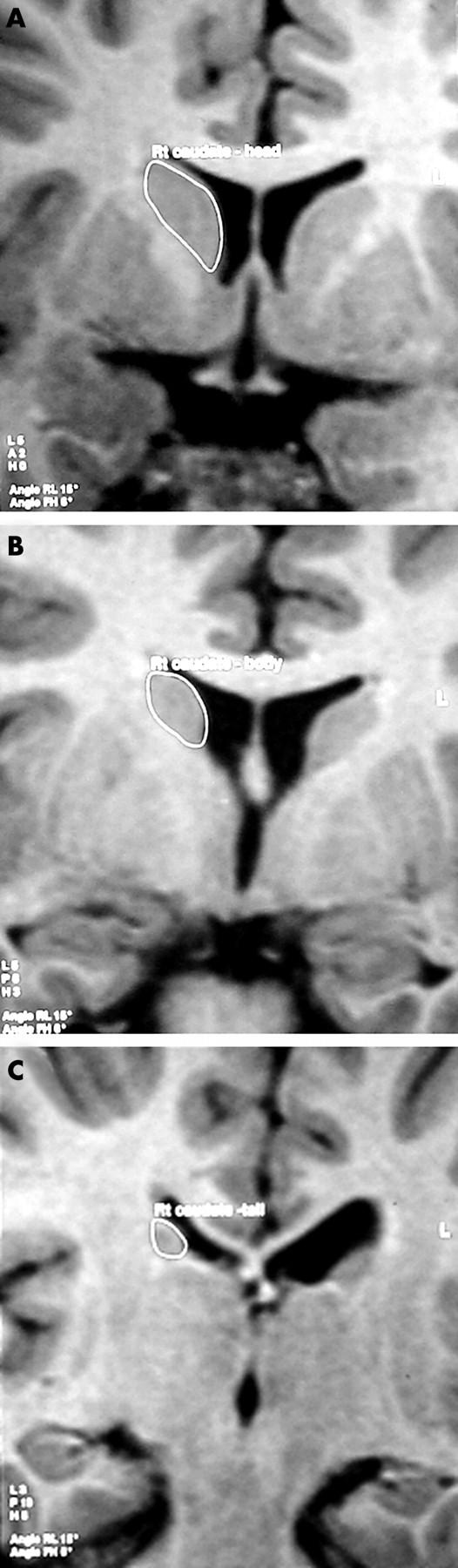
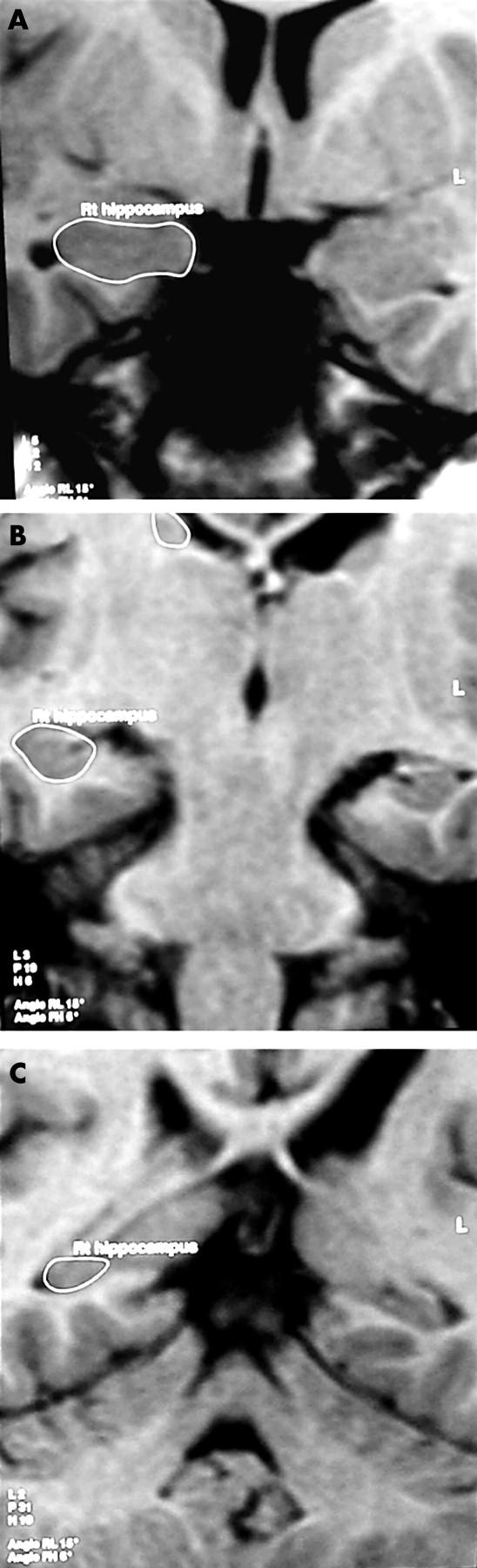
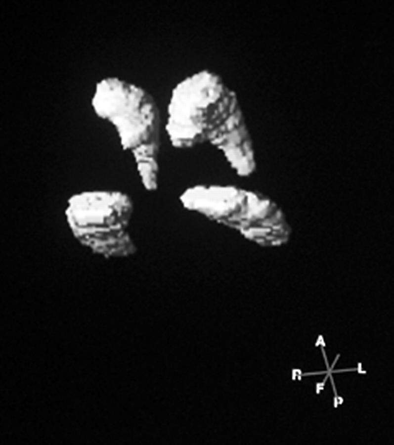
Methods: A total of 87 children (aged 15-16 years) with a history of VLBW (<1500 g) and eight age matched full term controls have been studied with detailed magnetic resonance brain scans. Volume measurements of the caudate nuclei and hippocampal formations were made.
Results: Scans in 42.5% of the children showed evidence of perinatal brain injury. There was no significant difference in IQ, dyspraxia, or attention deficit between children with qualitatively normal and abnormal scans. However, quantitative volumetric analysis showed that children with a low IQ had smaller volume measurements for the right caudate nucleus and left hippocampus, and a smaller hippocampal ratio (left volume:right volume) than those with normal IQ.
Conclusion: Data suggest that learning disorder, attention deficit, and dyspraxia in children who survive VLBW do not correlate with conventional markers of perinatal brain injury, and may be related to global brain growth and the development of key structures, such as the caudate nuclei and hippocampal formations.
Figures
References
Publication types
MeSH terms
LinkOut - more resources
Full Text Sources
Medical