. 2002 Oct 1;99(20):13166-71.
doi: 10.1073/pnas.152456799. Epub 2002 Sep 23.

Minimum molecular architectures for transcription and replication of the influenza virus

Affiliations

Minimum molecular architectures for transcription and replication of the influenza virus

Ayae Honda et al. Proc Natl Acad Sci U S A. .

Abstract

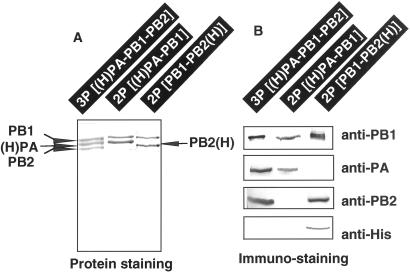
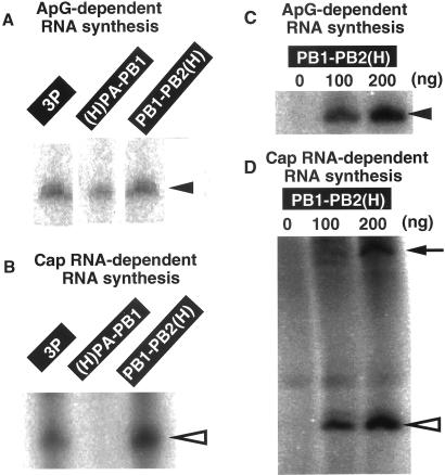
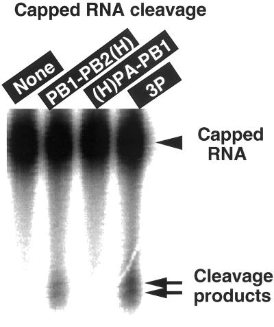
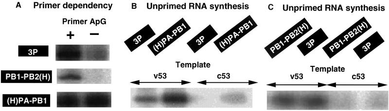
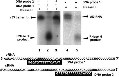
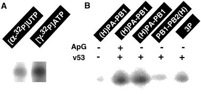
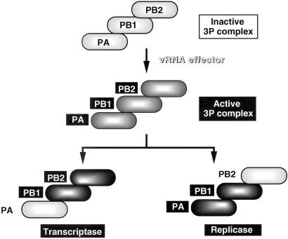
The RNA-dependent RNA polymerase of influenza virus is composed of three viral P proteins (PB1, PB2, and PA) and involved in both transcription and replication of the RNA genome. The PB1 subunit plays a key role in both the assembly of three P protein subunits and the catalytic function of RNA polymerization. We have established a simultaneous expression system of three P proteins in various combinations using recombinant baculoviruses, and isolated the PA-PB1-PB2 ternary (3P) complex and two kinds of the binary (2P) complex, PA-PB1 and PB1-PB2. The affinity-purified 3P complex showed all of the catalytic properties characteristic of the transcriptase, including capped RNA-binding, capped RNA cleavage, model viral RNA binding, model viral RNA-directed RNA synthesis, and polyadenylation of newly synthesized RNA. The PB1-PB2 binary complex showed essentially the same catalytic properties as does the 3P complex, whereas the PA-PB1 complex catalyzed de novo initiation of RNA synthesis in the absence of primers. Taken together we propose that the catalytic specificity of PB1 subunit is modulated to the transcriptase by binding PB2 or the replicase by interaction with PA.


Figures

Figure 1
Figure 1
Expression and purification of influenza virus P proteins. Three kinds of recombinant viruses, RBVPB1, RBVPB2 ,and RBVH-PA, were coinfected in various combinations onto Tn5 cells at multiplicity of infection of 2 for each virus (from left to right: RBVPB1 + RBVPB2 + RBVH-PA; RBVH-PA + RBVPB1; RHVPB1 + RBVPB2-H). After 4 days culture, the cells were harvested and the cell lysates were prepared as described in Materials and Methods. Whole cell lysates were fractionated into the supernatant (cytoplasm) and pellet (nuclei) by centrifugation for 5 min at 400 × g. The supernatants were subjected to Ni2+-ATM affinity purification as described in Materials and Methods. Imidazole elution fractions were pooled and subjected to SDS/PAGE. The gels were stained with CBB (A). The gels were also subjected to immunoblotting using anti-PB1, anti-PB2, anti-PA and anti-His tag antibodies (B). (H)PA represents the PA with the long His-tag at its N terminus, whereas PB2(H) represents the PB2 with the short His-tag at its C terminus. Anti-His-6 used in this study crossreacts with the short His-tag but not with the long His-tag.
Figure 2
Figure 2
Model vRNA-directed primer-dependent RNA synthesis in vitro by the 3P or 2P complexes. (A) RNA synthesis in vitro by the indicated P complexes [3P, 300 ng; (H)PA–PB1, 200 ng; PB1–PB2(H), 200 ng] was carried out using v53 template and in the presence of ApG primer. The conditions for in vitro RNA synthesis are as described in Materials and Methods. Products were analyzed by urea/10% PAGE, and the gel was exposed to an x-ray film. Filled triangle on right shows the template-sized RNA product. (B) RNA synthesis by the purified 3P, (H)PA–PB1 and PB1–PB2(H) complexes was carried out under the same conditions except that globin mRNA was used as a primer in place of ApG. Products were analyzed by urea/10% PAGE. The transcript, indicated by open triangle on right, is longer by about 10 nucleotides than ApG-primed transcript. (C) v53-directed ApG-primed RNA synthesis was carried out using the indicated amounts of the purified PB1–PB2(H) complex. Products were analyzed by urea/10% PAGE. (D) v53-directed globin mRNA-primed RNA synthesis was carried out as in the case of C. Open triangle on right shows the product with the capped primer, which is about 10 nucleotide-longer than the template, while arrow shows products with poly(A) tails.
Figure 3
Figure 3
Capped RNA endonuclease activity of the purified 3P and 2P complexes. The purified PB1–PB2(H), (H)PA–PB1 or (H)PA–PB1–PB2 (3P) complex were incubated with capped poly(A) with 32P only at cap-1 structure in the presence of 1 pmol v53 RNA. The amounts of P protein complexes used were the same as those used in transcription assay (see Fig. 2). After incubation for 30 min at 30°C, the cleavage products were analyzed by urea/8% PAGE. The gel was exposed to x-ray film. The RNA cleavage activity was not detected in the absence of v53 RNA.
Figure 4
Figure 4
Unprimed RNA synthesis by the 3P and 2P complexes. (A) v53-directed RNA synthesis by the (H)PA–PB1–PB2 (3P) (300 ng), PB1–PB2(H) (200 ng) or (H)PA–PB1 (200 ng) complexes was carried out in the presence (+) or absence (−) of ApG primer. Products were analyzed by urea/10% PAGE, and the gel was exposed to a x-ray film. (B) v53- or c53-directed unprimed RNA synthesis was carried out using the (H)PA–PB1–PB2 (3P) (300 ng) or (H)PA–PB1(200 ng) complexes. Products were analyzed as above. (C) v53- or c-53-directed unprimed RNA synthesis was carried out using the PB1–PB2(H) (200 ng) or 3P (300 ng) complexes (note that the exposure time was longer for C than for B).
Figure 5
Figure 5
Analysis of transcripts formed in unprimed RNA synthesis. (Lanes 1–3) RNA synthesized by the (H)PA–PB1 complex in the absence of primers and using [α-32P]UTP as a labeled substrate was hybridized with plus-strand (probe 1) or minus-strand (probe 2) DNA probes. After treatment with RNase H, RNA was isolated and analyzed by urea/12% PAGE. The gel was exposed to an imaging plate, which was developed with BAS1000 (Fuji). (Lanes 4 and 5) Radioactive c53 RNA (plus-strand) was synthesized by using T7 RNA polymerase and subjected to one cycle of hybridization and RNase H treatment.
Figure 6
Figure 6
De novo initiation of RNA synthesis by the 3P and 2P complexes. (A) v53-directed unprimed RNA synthesis by the (H)PA–PB1 complex was carried in the presence of either [α-32P]UTP or [γ-32P]ATP. The specific radioactivity was about 10-fold higher for [γ-32P]ATP than [α-32P]UTP. (B) RNA synthesis by the same amounts of 3P, (H)PA–PB1, and PB1–PB2(H) with respect to v53-directed ApG-primed RNA synthesis activity was carried out under the standard reaction conditions except that [γ-32P]ATP was added in place of [α-32P]UTP, and ApG and v53 were depleted for the reactions indicated.
Figure 7
Figure 7
Functional specificity of the 2P complexes. The 3P complex formed in insect cells after coinfection of three recombinant baculoviruses is inactive in RNA synthesis, but is converted into the active form in the presence of vRNA effector (17). Here it is demonstrated that the PB1–PB2(H) binary complex carries the catalytic specificity of transcriptase, whereas the (H)A-PB1 complex has the specificity of replicase.

References

    1. Palese P, Kingsbury D W, editors. Genetics of Influenza Viruses. Vienna: Springer; 1983.
    1. Krug R M, Alonso-Caplen F V, Julkenun I, Katze M G. In: The Influenza Viruses. Krug R M, editor. New York: Plenum; 1989. pp. 89–152.
    1. Ishihama A. Biochimie. 1996;78:1097–1102. - PubMed
    1. Honda A, Ishihama A. Biol Chem. 1997;378:483–488. - PubMed
    1. Bouloy M, Plotch S J, Krug R M. Proc Natl Acad Sci USA. 1978;75:4886–4890. - PMC - PubMed

Publication types

LinkOut - more resources