. 2002 Oct 1;99(20):12979-84.
doi: 10.1073/pnas.202300099. Epub 2002 Sep 23.

Functional correction of adult mdx mouse muscle using gutted adenoviral vectors expressing full-length dystrophin

Affiliations

Functional correction of adult mdx mouse muscle using gutted adenoviral vectors expressing full-length dystrophin

Christiana DelloRusso et al. Proc Natl Acad Sci U S A. .

Abstract

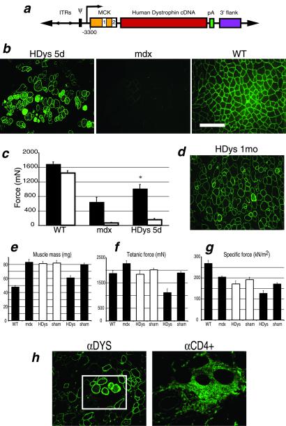
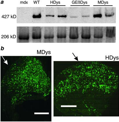
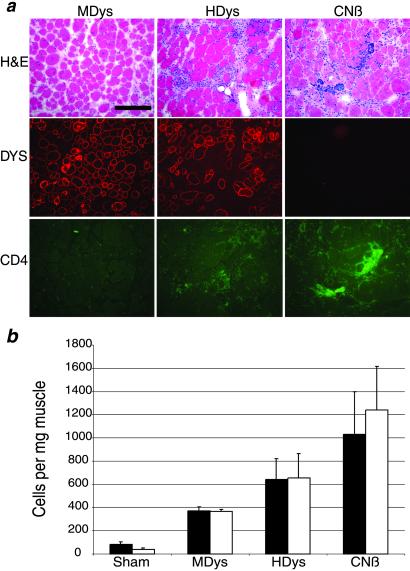
Duchenne muscular dystrophy is a lethal X-linked recessive disorder caused by mutations in the dystrophin gene. Delivery of functionally effective levels of dystrophin to immunocompetent, adult mdx (dystrophin-deficient) mice has been challenging because of the size of the gene, immune responses against viral vectors, and inefficient infection of mature muscle. Here we show that high titer stocks of three different gutted adenoviral vectors carrying full-length, muscle-specific, dystrophin expression cassettes are able to efficiently transduce muscles of 1-yr-old mdx mice. Single i.m. injections of viral vector restored dystrophin production to 25-30% of mouse limb muscle 1 mo after injection. Furthermore, functional tests of virally transduced muscles revealed almost 40% correction of their high susceptibility to contraction-induced injury. Our results show that functional abnormalities of dystrophic muscle can be corrected by delivery of full-length dystrophin to adult, immunocompetent mdx mice, raising the prospects for gene therapy of muscular dystrophies.


Figures

Figure 1
Figure 1
Analysis of mdx mouse muscles injected with HDys. (a) Plasmid vector used for generation of HDys gutted adenovirus: ITRs, inverted terminal repeats; Ψ, viral packaging signal; MCK gene regulatory element (the 6.5-kb MCK promoter/enhancer contains exon 1, intron 1, and a truncated exon 2) (8); 12.1-kb full-length human dystrophin cDNA; pA, simian virus 40 polyadenylation signal; and 3′-flank, 5.5-kb 3′-genomic flanking region of the human dystrophin gene. (b) Immunofluoresence (IF) analysis of cryosections from HDys-injected mdx, uninjected mdx, and C57BL/10J (WT) control mice. Anti-dystrophin Abs detected dystrophin expression localized to the sarcolemma (green). The scale bar applies to all IF photos and is equal to 100 μm. (c) TA muscles were analyzed in situ by measuring force production after two injury-inducing lengthening contractions, LC1 (filled bars) and LC2 (open bars). Muscles were tested 5 d after virus (HDys), or buffer (sham), injection into mdx mice. WT muscles were uninjected and age-matched. HDys-injected muscles showed significant recovery of force-producing capabilities after LC1 (P < 0.05 vs. mdx) and some protection after LC2. (d) After HDys injection (1 mo), IF analysis of TA muscles from mdx mice showed continuing high levels of dystrophin expression. However, eg demonstrate adverse physiological effects in the injected muscles not observed at 5 d (injected samples; open bars = 5 d, filled bars = 1 mo). HDys-injected muscles revealed decreases in mass and force significantly different from sham-injected and WT control muscles (P < 0.01). Each bar represents the mean of 6–8 muscles ± SEM. (h) IF inspection of HDys-injected muscles showed the presence of CD4+ (Right, green) and CD8+ (not shown) T cells surrounding some of the muscle fibers highly expressing dystrophin (green, enclosed in box); [×200 (Left) and boxed area magnified ×4 (Right)].
Figure 2
Figure 2
Mouse dystrophin protects against contraction-induced injury without adverse physiological consequences. (a) Plasmid constructs used to generate MDys and GEβDys gutted adenovirus. See Fig. 1 legend for abbreviations. MDys is identical to HDys with the exception of the species of the dystrophin cDNA. The promoter used in GEβDys is a 4.2-kb version of MCK that lacks the intron and exon 2 but contains an simian virus 40 viral protein 1 intron (9). (b) IF analysis of injected muscles show high levels of dystrophin expression 1 mo after injection. (Scale bar = 100 μm.) (c) Lengthening contractions in situ demonstrate significantly protected muscles 1 mo after injection of MDys gutted Ad. Protection is afforded after both LC1 (filled bars) and LC2 (open bars) (P < 0.05). GEβDys-injected muscles were protected after both contractions, but the effect was not significant. Control data from WT and mdx mice is the same as shown in Fig. 1c. (d) The protection against injury after virus injection was not accompanied by any loss in mass or force production. Control WT and mdx data are the same as shown in Fig. 1 eg. Bars represent the mean of 6–8 muscles ± SEM.
Figure 3
Figure 3
Widespread, high level expression of dystrophin in adult mdx mice 1 mo after injection with gutted Ad. (a) Immunoblot analysis of protein extracts from control and injected TA muscles shows varying levels of the full-length 427-kDa dystrophin protein. Loading controls are shown below in an identical Coomassie-stained gel. Myosin is seen here at 206 kDa. (b) Montage photos of entire cross sections of TA muscles from 1-yr-old mdx mice injected with MDys (Left) or HDys (Right) demonstrate widespread expression of dystrophin from the approximate site of injection (arrows) 1 mo later. (Scale bar = 1.0 mm.)
Figure 4
Figure 4
HDys induces less immune cell infiltration than a first-generation adenovirus expressing β-gal but more than MDys. (a) MDys-, HDys- and CNβ-injected muscle serial cryosections are shown stained with hematoxylin/eosin, anti-dystrophin antisera (Dys, red), or anti-CD4+ cell Ab (CD4+, green). Note the areas of mononuclear cell infiltration (hematoxylin/eosin) and CD4+ cells in the HDys- and CNβ- injected muscles. Results of flow cytometry analysis of injected muscles are shown in b. Filled bars = CD4+ cells; open bars = CD8+ cells. Each bar represents the mean of four muscles ± SEM. Mdx muscles injected with helper adenovirus at the levels present in our gutted Ad preparations contained the same number of infiltrating T cells as did sham-injected muscles (data not shown).

References

    1. Emery A E H. Duchenne Muscular Dystrophy. Oxford: Oxford Medical Publications; 1993.
    1. Miller R G, Hoffman E P. Neurol Clin. 1994;12:699–725. - PubMed
    1. Hoffman E P, Morgan J E, Watkins S C, Partridge T A. J Neurol Sci. 1990;99:9–25. - PubMed
    1. Sicinski P, Geng Y, Ryder-Cook A S, Barnard E A, Darlison M G, Barnard P J. Science. 1989;244:1578–1580. - PubMed
    1. Brooks S V. J Muscle Res Cell Motil. 1998;19:179–187. - PubMed

Publication types

LinkOut - more resources