Review
. 2002 Oct;130(1):4-11.
doi: 10.1046/j.1365-2249.2002.01953.x.

The dual function of the splenic marginal zone: essential for initiation of anti-TI-2 responses but also vital in the general first-line defense against blood-borne antigens

Affiliations
Review

The dual function of the splenic marginal zone: essential for initiation of anti-TI-2 responses but also vital in the general first-line defense against blood-borne antigens

A Zandvoort et al. Clin Exp Immunol. 2002 Oct.

Abstract

The splenic marginal zone (S-MZ) is especially well equipped for rapid humoral responses and is unique in its ability to initiate an immune response to encapsulated bacteria (T-cell independent type 2 (TI-2) antigens). Because of the rapid spreading through the blood, infections with blood-borne bacteria form a major health risk. To cope with blood-borne antigens, a system is needed that can respond rapidly to a great diversity of organisms. Because of a number of unique features, S-MZ B cells can respond rapid and efficient to all sorts of blood-borne antigens. These unique features include a low blood flow microenvironment, low threshold for activation, high expression of complement receptor 2 (CR2, CD21) and multireactivity. Because of the unique high expression of CD21 in a low flow compartment, S-MZ B cells can bind and respond to TI-2 antigens even with relatively low-avid B cell receptors. Although TI-2 antigens are in general poorly opsonized by classic opsonins, a particular characteristic of these antigens is their ability to bind very rapidly to complement fragment C3d without the necessity of previous immunoglobulin binding. TI-2 primed S-MZ B cells, already by first passage through the germinal centre, will meet antigen-C3d complexes bound to follicular dendritic cells, allowing unique immediate isotype switching. This explains that the primary humoral response to TI-2 antigens is unique in its characterization by a rapid increase in IgM concurrent with IgG antibody levels.


Figures
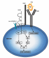
Fig. 1
Fig. 1
The CD21, CD19, TAPA-1, and Leu-13membrane complex expressed on B cells modulates the BCR signalling. After binding of complement-opsonized (TI-2) antigen to the BCR, an intracellular signal cascade is activated starting with the activation of several tyrosine kinases like fyn, blk, lyn and syk. These tyrosine kinases can activated Btk. Activation of Btk is also dependent on phosphatidylinositol 3-kinase (PI-3K), which is recruited via phosphorylation of intracytoplasmic residues of CD19. Btk induces a variety of downstream effector mechanisms like calcium signalling via phospholipase Cγ (PLCγ) and inositol 3,4,5-triphosphate (IP-3). Through activation of CD21 by complement fragment C3d, CD19 is activated. CD21 and CD19 interact through their extracellular and transmembrane region. CD19 and TAPA-1/Leu-13 are associated through their extracellular region. This results in the activation of Vav which, via a number of downstream processes and phosphatidylinositol 4-phosphate 5-kinase (PIP-5K), also mediated calcium signalling through IP-3. Based on [23,9,24,29].
Fig. 2
Fig. 2
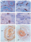
(a–d) Localization pattern of PPS in the rat spleen. Rats were injected intravenously with PPS and sacrificed at (a) 15 min, (b) 1 day, or (c) 5 days after treatment. PPS localization was demonstrated with an immunohistochemical method using a polyclonal antibody directed to PPS type 19. 15 min after treatment, PPS localizes extensively in the S-MZ (a), 1 day after injection, PPS is detected in the primary follicle and 5 days after vaccination, PPS binding is observed in a FDC like pattern in the follicles. (d) No binding is observed in animals, decomplemented by cobra venom factor (CVF) treatment. S-MZ, marginal zone; P, periarteriolar lymphoid sheath; RP, red pulp; F, follicle. (e–f): Localization pattern of PPS (e) type 2 and (f) type 9 N in the human spleen. Spleen sections were incubated with PPS in combination with serum (complement). Sections were subsequently incubated with type specific antibodies against PPS. Strong PPS staining on FDC's in the germinal centres and on B cells in the S-MZ. S-MZ, marginal zone; RP, red pulp; Cor, corona; GC, germinal centre.
Fig. 3
Fig. 3
Immune responses in rats after Tetanus Toxoid (TT) or PPS vaccination. Rats were intravenously vaccinated with either TT (Tetavax®) or PPS (Pneumovax ®) and blood was sampled at different time points after vaccination. A simultaneous, rapid IgM (▪) and IgG (d) response was observed after PPS vaccination, which declined to almost prevaccination values at day 24. Rats vaccinated with TT showed a late induction of only IgG antibodies (▴). IgM antibodies after TT vaccination were hardly detected. Values represent the average of 6–9 rats. Antibody titres are depicted in U/ml.
Fig. 4
Fig. 4
Migration pattern of antigen activated marginal zone B cells. Naïve B cells enter the marginal zone through the marginal sinus. After antigenic stimulation, activated B cells can migrate directly to the red pulp (A) to accumulate in extra follicular zones and differentiate to antibody producing plasma cells (B) or migrate to the PALS (C) were they can accumulate in the outer PALS (D) followed by clonal expansion. Activated cells can migrate to the red pulp through bridging channels (exit zones) (E). Activated B cells can also migrate to the primary follicle (F) to initiate a germinal centre process with clonal expansion and affinity maturation (G). In the germinal centre, memory B cell are generated which can directly colonize the marginal zone (H). After TI-2 antigenic stimulation, only few activated cells will initiate a germinal centre process. S-MZ, splenic marginal zone; PALS, periarteriolar lymphoid sheath; Cor, lymphocyte corona; GC, germinal centre; RP, red pulp.

References

    1. Martin F, Oliver AM, Kearney JF. Marginal zone and B1 B cells unite in the early response against T-independent blood-borne particulate antigens. Immunity. 2001;14:617–29. - PubMed
    1. Timens W, Boes A, Poppema S. Human marginal zone B cells are not an activated B cell subset. strong expression of CD21 as a putative mediator for rapid B cell activation. Eur J Immunol. 1989;19:2163–6. - PubMed
    1. Harms G, Hardonk MJ, Timens W. In vitro complement-dependent binding and in vivo kinetics of pneumococcal polysaccharide TI-2 antigens in the rat spleen marginal zone and follicle. Infect Immun. 1996;64:4220–5. - PMC - PubMed
    1. Guinamard R, Okigaki M, Schlessinger J, Ravetch JV. Absence of marginal zone B cells in Pyk-2-deficient mice defines their role in the humoral response. Nat Immunol. 2000;1:31–6. - PubMed
    1. Kraal G. Cells in the marginal zone of the spleen. Int Rev Cytol. 1992;132:31–74. - PubMed

Publication types

MeSH terms