. 2002 Oct 1;99(20):12645-50.
doi: 10.1073/pnas.202476799. Epub 2002 Sep 24.

Crystallographic findings on the internally uncoupled and near-rigor states of myosin: further insights into the mechanics of the motor

Affiliations

Crystallographic findings on the internally uncoupled and near-rigor states of myosin: further insights into the mechanics of the motor

D M Himmel et al. Proc Natl Acad Sci U S A. .

Abstract

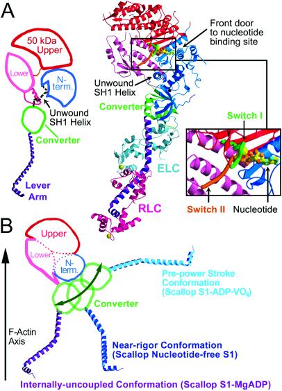
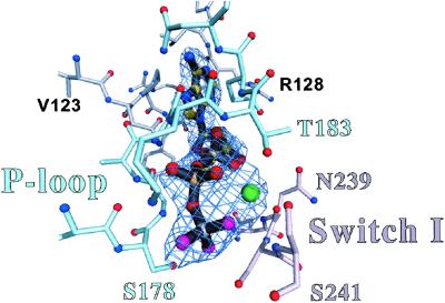
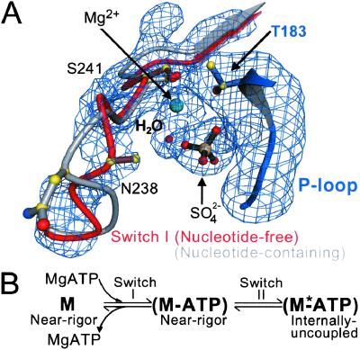
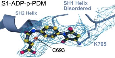
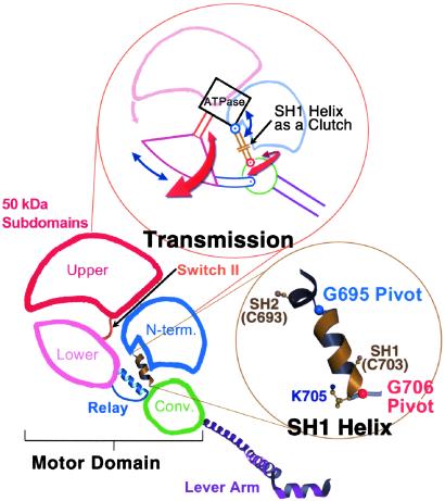
Here we report a 2.3-A crystal structure of scallop myosin S1 complexed with ADP.BeF(x), as well as three additional structures (at 2.8-3.8 A resolution) for this S1 complexed with ATP analogs, some of which are cross-linked by para-phenyl dimaleimide, a short intramolecular cross-linker. In all cases, the complexes are characterized by an unwound SH1 helix first seen in an unusual 2.5-A scallop myosin-MgADP structure and described as corresponding to a previously unrecognized actin-detached internally uncoupled state. The unwinding of the SH1 helix effectively uncouples the converter/lever arm module from the motor and allows cross-linking by para-phenyl dimaleimide, which has been shown to occur only in weak actin-binding states of the molecule. Mutations near the metastable SH1 helix that disable the motor can be accounted for by viewing this structural element as a clutch controlling the transmission of torque to the lever arm. We have also determined a 3.2-A nucleotide-free structure of scallop myosin S1, which suggests that in the near-rigor state there are two conformations in the switch I loop, depending on whether nucleotide is present. Analysis of the subdomain motions in the weak actin-binding states revealed by x-ray crystallography, together with recent electron microscopic results, clarify the mechanical roles of the parts of the motor in the course of the contractile cycle and suggest how strong binding to actin triggers both the power stroke and product release.


Figures

Figure 1
Figure 1
Overview of the internally uncoupled scallop S1 conformation. (A) (Center) The internally uncoupled S1-ADP⋅BeFx structure (50-kDa upper subdomain is shown in red, 50-kDa lower subdomain is shown in pink, N-terminal subdomain is shown in blue, converter is shown in green, lever arm heavy chain is shown in purple, essential light chain is shown in cyan, regulatory light chain is shown in magenta). Notable features of the structure include the unusual position of the lever arm and the unwound SH1 helix. Three other scallop structures (S1-AMPPNP, S1-ATP[γ-S]-p-PDM, and S1-ADP-p-PDM), as well as the previously reported scallop S1-MgADP (1), show the same conformation, although the orientations of the respective lever arms vary slightly. (Right Inset) Expanded view of the nucleotide binding size. (Left) Schematic diagram of the internally uncoupled conformation, showing the subdomains and the approximate location of the disordered SH1 helix (light chains not shown). (B) For comparison, a two-dimensional projection of the three weak actin-binding S1 conformations observed in scallop crystal structures (light chains not shown) (1, 2). The 50-kDa upper and N-terminal subdomains of the three structures are superimposed to show the large changes in the relative positions of the converter and lever arm (see also ref. 2). In each conformation, the 50-kDa lower subdomain adopts a slightly different position with respect to the 50-kDa upper and N-terminal subdomains, leading to a markedly different orientation of the converter. The converter, in turn, controls the position of the lever arm (1). Movement of the 50-kDa lower subdomain is not represented in this schematic diagram. This subdomain rotates to maintain contact with the converter as S1 adopts each of the three conformations. See Fig. 5 for a more detailed three-dimensional description of the subdomain motions.
Figure 2
Figure 2
Nucleotide electron density. The nucleotide binding site is shown together with a simulated annealing FoFc omit map of ADP⋅BeFx, contoured at the 5.0 σ level. The P loop (part of the N-terminal subdomain) is shown in sky-blue, and switch I (lower right, part of the 50-kDa upper subdomain) is shown in pink. Included in this view is the electron density attributed to the Mg2+ ion (green ball). The γ-phosphate position is occupied by BeFx (Be in gray and F in magenta). The other internally uncoupled structures show approximately the same nucleotide conformation, but at a lower resolution. The interactions of the Mg2+ nucleotide with the P loop and switch I are virtually identical to those previously reported at higher resolution in a near-rigor Dictyostelium truncated myosin-ATP structure (8).
Figure 3
Figure 3
A conformational change in switch I on the binding of nucleotide. (A) Shown is a composite of four difference Fourier (FoFc) electron density omit maps of switch I and the surrounding region of the nucleotide-binding site of the nucleotide-free near-rigor structure. Each map was generated by torsional simulated-annealing in which atoms were omitted either for a segment of switch I (residues N235 to R242), the Mg2+ ion and a water molecule, a sulfate ion, or part of the P loop (residues A180 to T183). The maps are contoured at the 3.5 σ level. Superimposed on the maps is the proposed conformational change of switch I in the transition from the nucleotide-free to the nucleotide-bound (ATP and its analogs, or ADP) states. The P loop of the scallop near-rigor and S1-ADP⋅BeFx structures are superimposed. The “empty” nucleotide-binding site of the near-rigor conformation is shown in color (switch I in red, P loop in blue, Mg2+ ion in green, with a sulfate ion occupying the β-phosphate position). In gray is the same site in the internally uncoupled scallop S1-ADP⋅BeFx structure (nucleotide not shown), which shows the conformation of switch I seen in all nucleotide-containing myosin crystal structures. When nucleotide is present in the binding site, it appears to displace part of switch I, most notably residue N238. The internally uncoupled state is initiated by small changes in the switch II loop (not shown, see ref. 1). (B) The equilibrium equation (in vitro) between the empty and nucleotide-containing near-rigor myosin structures, leading to the internally uncoupled state. Shown also are the steps at which switches I and II change. An asterisk is assigned to the internally uncoupled ATP state (right) to indicate that this conformation may correspond to the second ATP state predicted in biochemical kinetics studies (19). The near-rigor nucleotide-free state (left) is not likely to be part of the in vivo acto-myosin contractile cycle.
Figure 4
Figure 4
Electron density for the cross-linker. Shown is the simulated annealing FoFc omit map for the electron density of p-PDM together with the cross-linker modeled into the structure of S1-ADP-p-PDM. To generate this map, contoured at the 3.0 σ level, residues C693 and K705 were omitted along with the p-PDM model. The SH1 helix is disordered, including the SH1 sulfhydryl (C703). Biochemical studies with rabbit muscle myosin have indicated that this thiol can be cross-linked to the SH2 sulfhydryl (C693) by p-PDM (15, 24, 25). However, this map reveals that, in scallop S1, p-PDM cross-links the SH2 sulfhydryl to the side chain of K705, instead of SH1. The scallop S1-ATP[γ-S]-p-PDM structure gives the same result.
Figure 5
Figure 5
Mechanical components of the motor. Schematic diagram of the myosin head (shown here in the near-rigor conformation), depicting the four subdomains of the motor together with the lever arm (light chains not shown). (Upper Inset) a mechanical representation of the motor's “transmission,” illustrated to highlight the roles of various components. Switch II functions like a piston by delivering work from the enzyme (“ATPase”) to the 50-kDa lower subdomain, which converts this work to rotational motion, roughly comparable to the role of a crank shaft. During the transition from the near-rigor to the pre-power stroke conformation (with the lever arm moving from “down” to “up”), the rotation of the 50-kDa lower subdomain can be divided into two roughly perpendicular vector components (red and blue arrows). The red arrows represent rotation about axes in the plane of the paper, and the blue arrows represent rotation about axes perpendicular to the plane of the paper (described previously as twist and tilt motions, respectively; ref. 40). The converter rotates about the glycine pivots along these vector components as shown (1, 10) and, in addition moves about a hinge in the relay (not shown, see text and ref. 1). During this transition, the molecule passes through the internally uncoupled state, in which the SH1 helix unwinds. The converter/lever arm orientation then becomes less constrained, and torque cannot be delivered from switch II to the converter/lever arm module. Thus, the function of the SH1 helix can be compared with that of an automobile clutch, which can disengage the gear train and drive shaft from the motor. Based on EM studies of myosin cross-bridges bound to actin and the x-ray crystallographic nucleotide-free near-rigor structure, it appears that rotation of the 50-kDa upper subdomain (pink arrow) on strong binding to actin closes the front of the cleft and triggers both the power stroke and product release (see text). A related two-dimensional mechanical model of the myosin motor has been published (41), but revisions are needed to agree with recent experimental findings. (Lower Inset) The SH1 helix flanked by the glycine pivots.

References

    1. Houdusse A, Kalabokis V N, Himmel D, Szent-Györgyi A G, Cohen C. Cell. 1999;97:459–470. - PubMed
    1. Houdusse A, Szent-Györgyi A G, Cohen C. Proc Natl Acad Sci USA. 2000;97:11238–11243. - PMC - PubMed
    1. Goldman Y E. Cell. 1998;93:1–4. - PubMed
    1. Spudich J A. Nature (London) 1994;372:515–518. - PubMed
    1. Lymn R W, Taylor E W. Biochemistry. 1971;10:4617–4624. - PubMed

Publication types

LinkOut - more resources