. 2002 Oct;68(10):4906-14.
doi: 10.1128/AEM.68.10.4906-4914.2002.

Diversity of endophytic bacterial populations and their interaction with Xylella fastidiosa in citrus plants

Affiliations

Diversity of endophytic bacterial populations and their interaction with Xylella fastidiosa in citrus plants

Welington L Araújo et al. Appl Environ Microbiol. 2002 Oct.

Abstract

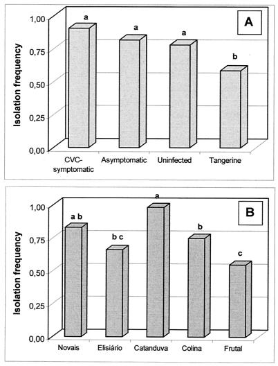
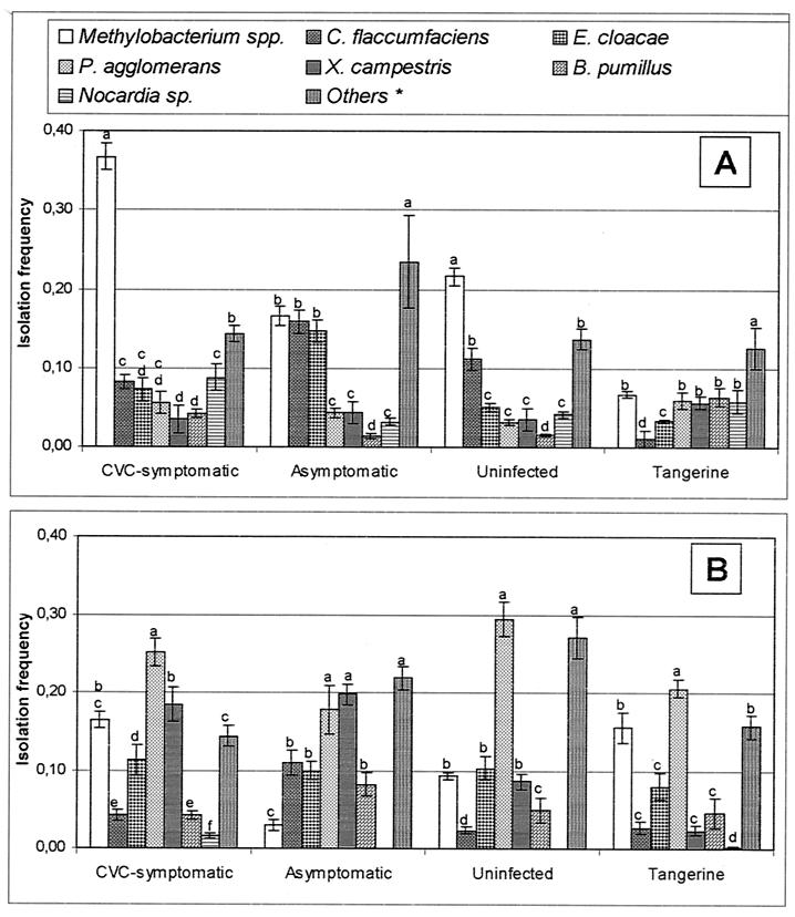
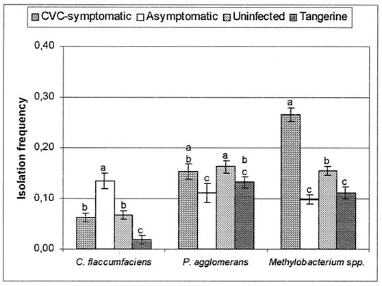
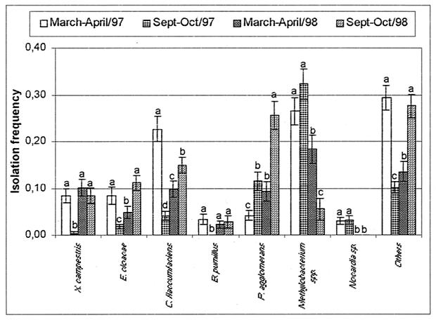
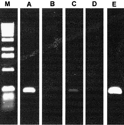
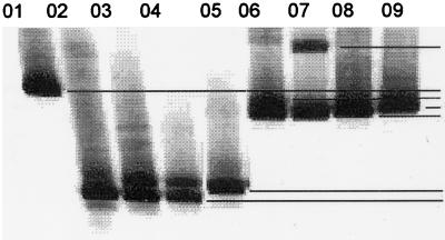
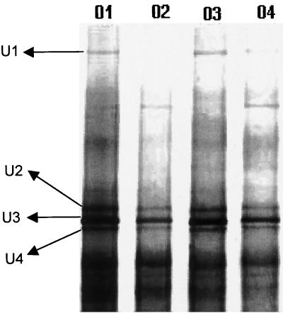
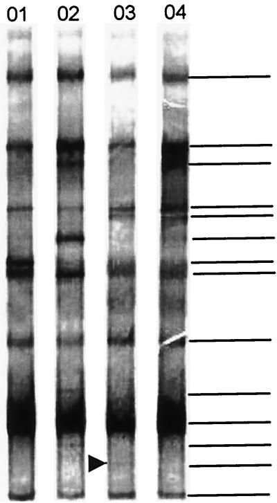
Citrus variegated chlorosis (CVC) is caused by Xylella fastidiosa, a phytopathogenic bacterium that can infect all Citrus sinensis cultivars. The endophytic bacterial communities of healthy, resistant, and CVC-affected citrus plants were studied by using cultivation as well as cultivation-independent techniques. The endophytic communities were assessed in surface-disinfected citrus branches by plating and denaturing gradient gel electrophoresis (DGGE). Dominant isolates were characterized by fatty-acid methyl ester analysis as Bacillus pumilus, Curtobacterium flaccumfaciens, Enterobacter cloacae, Methylobacterium spp. (including Methylobacterium extorquens, M. fujisawaense, M. mesophilicum, M. radiotolerans, and M. zatmanii), Nocardia sp., Pantoea agglomerans, and Xanthomonas campestris. We observed a relationship between CVC symptoms and the frequency of isolation of species of Methylobacterium, the genus that we most frequently isolated from symptomatic plants. In contrast, we isolated C. flaccumfaciens significantly more frequently from asymptomatic plants than from those with symptoms of CVC while P. agglomerans was frequently isolated from tangerine (Citrus reticulata) and sweet-orange (C. sinensis) plants, irrespective of whether the plants were symptomatic or asymptomatic or showed symptoms of CVC. DGGE analysis of 16S rRNA gene fragments amplified from total plant DNA resulted in several bands that matched those from the bacterial isolates, indicating that DGGE profiles can be used to detect some endophytic bacteria of citrus plants. However, some bands had no match with any isolate, suggesting the occurrence of other, nonculturable or as yet uncultured, endophytic bacteria. A specific band with a high G+C ratio was observed only in asymptomatic plants. The higher frequency of C. flaccumfaciens in asymptomatic plants suggests a role for this organism in the resistance of plants to CVC.


Figures

FIG. 1.
FIG. 1.
IFs of citrus endophytic bacteria from different citrus plants (A) from five citrus-growing areas (B). Means with the same letter are not significantly different by Duncan's test (P > 0.05).
FIG. 2.
FIG. 2.
IFs of endophytic bacteria from citrus plants in two sampling periods, March and April (A) and September and October (B) of 1997. Means within a plant category with the same letter are not significantly different by Duncan's test (P > 0.05). *, Other isolates included are Bacillus cereus, Burkholderia cepacia, Streptomyces sp., and unidentified isolates.
FIG. 3.
FIG. 3.
IFs of C. flaccumfaciens, P. agglomerans, and Methylobacterium spp. in branches of CVC-symptomatic, asymptomatic, uninfected, and tangerine plants. Means within a bacterial group with the same letter are not significantly different by Duncan's test (P > 0.05). Error bars indicate standard errors of the means (at least 40 plants were sampled).
FIG. 4.
FIG. 4.
IFs of endophytic bacteria from peeled citrus branches sampled in March and April and in September and October of 1997 and 1998 in the Brazilian city of Novais, São Paulo. Means within a bacterial group with the same letter are not significantly different by Duncan's test (P > 0.05). Error bars indicate standard errors of the means (at least 16 plants were sampled).
FIG. 5.
FIG. 5.
Detection of X. fastidiosa in citrus plants with specific primers for CVC-causing strains. All plants studied were evaluated for the presence of X. fastidiosa. Lanes: M, molecular size marker (Life Technologies); A, CVC-symptomatic plants; B, uninfected plants; C, asymptomatic plants; D, tangerine plants; E, positive control containing X. fastidiosa DNA.
FIG. 6.
FIG. 6.
DGGE profile of Methylobacterium spp. isolated from the branches of citrus plants. Lanes: 01, M. mesophilicum haplotype H (AR3/19); 02, Methylobacterium sp. haplotype G (AR1.6/4); 03, M. fujisawaense haplotype F (PR5/4); 04, M. fujisawaense haplotype F (PR5.1/1); 05, M. extorquens haplotype E (AR1.6/11); 06, M. fujisawaense haplotype D (SR5/3); 07, M. zatmanii haplotype C (SR1.6/2); 08, M. mesophilicum haplotype B (SR1.6/6); 09, M. zatmanii haplotype B (PR3/8). The codes in parentheses indicate the strains used.
FIG. 7.
FIG. 7.
Fingerprinting of the bacterial endophytic community of citrus branches by DGGE separation and analysis of 16S rDNA fragments amplified with the bacterial primer set 968F (primer F968⊥GC without GC clamp) plus 1387R. Lanes: 01, CVC-symptomatic plants; 02, healthy plants; 03, resistant plants; 04, tangerine plants. Bands U1, U2, U3, and U4 were excised and sequenced, and the sequence was shown to have between 92 and 98% sequence similarity to Nocardia nova (98%), Methylobacterium sp. (99%), Curtobacterium sp. (92%), and Corynebacterium accolens (99%).
FIG. 8.
FIG. 8.
DGGE analysis of 16S rDNA fragments of citrus plant samples collected in the Brazilian town of Novais, São Paulo. Lanes: 01, tangerine plants; 02, CVC-symptomatic plants; 03, asymptomatic plants; 04, uninfected plants. The 16S rDNA genes were previously amplified with specific primers for β-proteobacteria and then with bacterial universal primers with a GC clamp. The band with a high percentage of G+C was observed in all of the asymptomatic plants (arrow) but not in susceptible plants. The horizontal lines indicate the bands used for analysis. At least 20 plants within each category were used.

References

    1. Abdoulaye, S. Y., E. Giraud, P. Jourand, N. Garcia, A. Willems, P. de Lajudie, Y. Prin, M. Neyra, M. Gillis, C. Bivin-Masson, and B. Dreyfus. 2001. Methylotrophic Methylobacterium bacteria nodulate and fix nitrogen in symbiosis with legumes. J. Bacteriol. 183:214-220. - PMC - PubMed
    1. Araújo, W. L., H. O. Saridakis, P. A. V. Barroso, C. I. Aguilar-Vildoso, and J. L. Azevedo. 2001. Variability and interactions between endophytic bacteria and fungi isolated from leaf tissues of citrus rootstocks. Can. J. Microbiol. 47:229-236. - PubMed
    1. Azevedo, J. L., W. Maccheroni, Jr., J. O. Pereira, and W. L. Araújo. 15April2000, posting date. Endophytic microorganisms: a review on insect control and recent advances on tropical plants. Bio/Technology 3:40-65. [Online.] http://www.ejb.org/content/vol3/issue1/full/3/4.
    1. Bent, E., and C. P. Chanway. 1998. The growth-promoting effects of a bacterial endophyte on lodgepole pine are partially inhibited by the presence of other rhizobacteria. Can. J. Microbiol. 44:980-988.
    1. Chang, C. J., M. Garnier, L. Zreik, V. Rossetti, and J. M. Bove. 1993. Culture and serological detection of the xylem-limited bacterium causing citrus variegated chlorosis and its identification as a strain of Xylella fastidiosa. Curr. Microbiol. 27:137-142. - PubMed

Publication types

MeSH terms

LinkOut - more resources