

Double-staining method for differentiation of morphological changes and membrane integrity of Campylobacter coli cells
- PMID: 12324366
- PMCID: PMC126449
- DOI: 10.1128/AEM.68.10.5151-5154.2002
Double-staining method for differentiation of morphological changes and membrane integrity of Campylobacter coli cells
Abstract
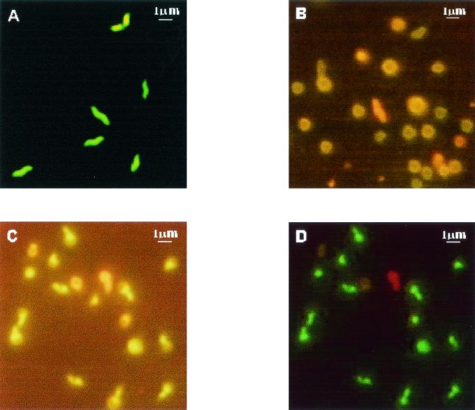
We developed a double-staining procedure involving NanoOrange dye (Molecular Probes, Eugene, Oreg.) and membrane integrity stains (LIVE/DEAD BacLight kit; Molecular Probes) to show the morphological and membrane integrity changes of Campylobacter coli cells during growth. The conversion from a spiral to a coccoid morphology via intermediary forms and the membrane integrity changes of the C. coli cells can be detected with the double-staining procedure. Our data indicate that young or actively growing cells are mainly spiral shaped (green-stained cells), but older cells undergo a degenerative change to coccoid forms (red-stained cells). Club-shaped transition cell forms were observed with NanoOrange stain. Chlorinated drinking water affected the viability but not the morphology of C. coli cells.
Figures
References
-
- American Public Health Association. 1995. Standard methods for the examination of water and wastewater, 19th ed. American Public Health Association, Washington, D.C.
-
- Colwell, R. R., and D. J. Grimes. 2000. Semantics and strategies, p. 1-6. In I. Nachamkin and M. J. Blaser (ed.), Campylobacter, 2nd ed. American Society for Microbiology, Washington, D.C.
-
- Griffiths, P. L. 1993. Morphological changes of Campylobacter jejuni growing in liquid culture. Lett. Appl. Microbiol. 17:152-155. - PubMed
-
- Harvey, P., and S. Leach. 1998. Analysis of coccal cell formation by Campylobacter jejuni using continuous culture techniques, and the importance of oxidative stress. J. Appl. Microbiol. 85:398-404. - PubMed
Publication types
MeSH terms
Substances
LinkOut - more resources
Full Text Sources
Molecular Biology Databases
