

Prediction of structure and function of G protein-coupled receptors
- PMID: 12351677
- PMCID: PMC130510
- DOI: 10.1073/pnas.122357199
Prediction of structure and function of G protein-coupled receptors
Abstract
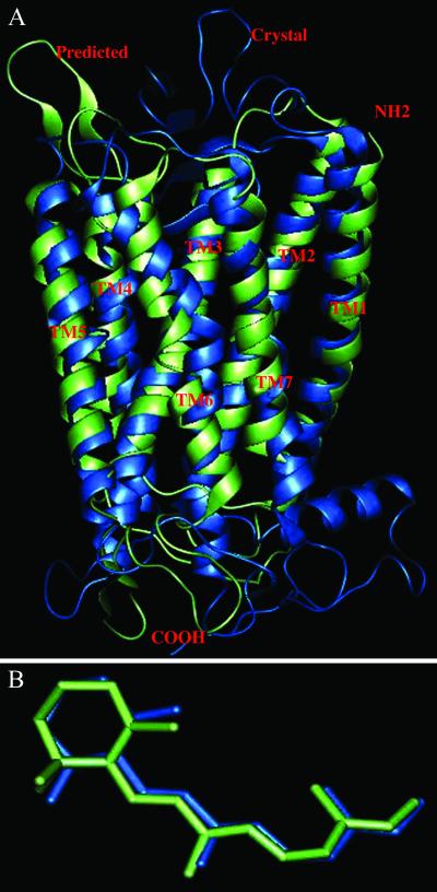
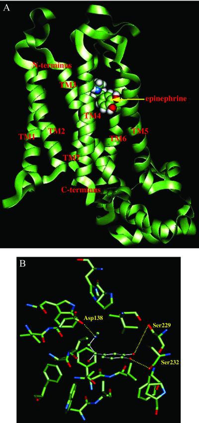
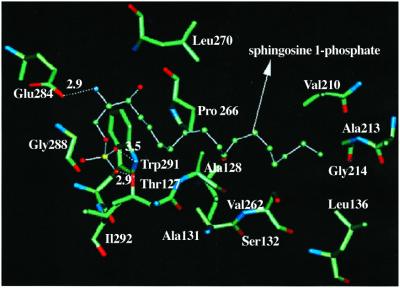
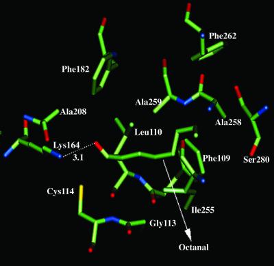
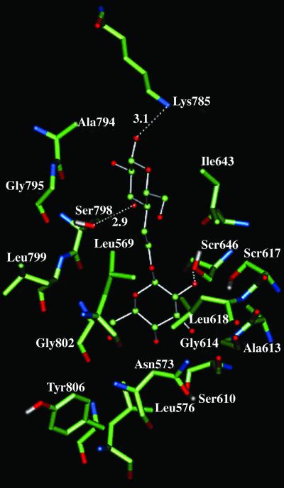
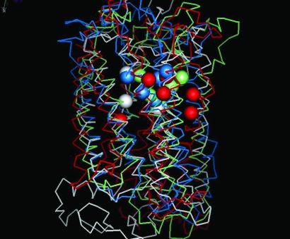
G protein-coupled receptors (GPCRs) mediate our sense of vision, smell, taste, and pain. They are also involved in cell recognition and communication processes, and hence have emerged as a prominent superfamily for drug targets. Unfortunately, the atomic-level structure is available for only one GPCR (bovine rhodopsin), making it difficult to use structure-based methods to design drugs and mutation experiments. We have recently developed first principles methods (MembStruk and HierDock) for predicting structure of GPCRs, and for predicting the ligand binding sites and relative binding affinities. Comparing to the one case with structural data, bovine rhodopsin, we find good accuracy in both the structure of the protein and of the bound ligand. We report here the application of MembStruk and HierDock to beta1-adrenergic receptor, endothelial differential gene 6, mouse and rat I7 olfactory receptors, and human sweet receptor. We find that the predicted structure of beta1-adrenergic receptor leads to a binding site for epinephrine that agrees well with the mutation experiments. Similarly the predicted binding sites and affinities for endothelial differential gene 6, mouse and rat I7 olfactory receptors, and human sweet receptor are consistent with the available experimental data. These predicted structures and binding sites allow the design of mutation experiments to validate and improve the structure and function prediction methods. As these structures are validated they can be used as targets for the design of new receptor-selective antagonists or agonists for GPCRs.
Figures
Similar articles
-
First principles predictions of the structure and function of g-protein-coupled receptors: validation for bovine rhodopsin.Biophys J. 2004 Apr;86(4):1904-21. doi: 10.1016/S0006-3495(04)74256-3. Biophys J. 2004. PMID: 15041637 Free PMC article.
-
Docking Simulations of G-Protein Coupled Receptors Uncover Crossover Binding Patterns of Diverse Ligands to Angiotensin, Alpha-Adrenergic and Opioid Receptors: Implications for Cardiovascular Disease and Addiction.Biomolecules. 2025 Jun 11;15(6):855. doi: 10.3390/biom15060855. Biomolecules. 2025. PMID: 40563495 Free PMC article.
-
Predicted 3D structure for the human beta 2 adrenergic receptor and its binding site for agonists and antagonists.Proc Natl Acad Sci U S A. 2004 Mar 2;101(9):2736-41. doi: 10.1073/pnas.0308751101. Epub 2004 Feb 23. Proc Natl Acad Sci U S A. 2004. PMID: 14981238 Free PMC article.
-
The Black Book of Psychotropic Dosing and Monitoring.Psychopharmacol Bull. 2024 Jul 8;54(3):8-59. Psychopharmacol Bull. 2024. PMID: 38993656 Free PMC article. Review.
-
Factors that impact on the use of mechanical ventilation weaning protocols in critically ill adults and children: a qualitative evidence-synthesis.Cochrane Database Syst Rev. 2016 Oct 4;10(10):CD011812. doi: 10.1002/14651858.CD011812.pub2. Cochrane Database Syst Rev. 2016. PMID: 27699783 Free PMC article.
Cited by
-
Predicted structure of agonist-bound glucagon-like peptide 1 receptor, a class B G protein-coupled receptor.Proc Natl Acad Sci U S A. 2012 Dec 4;109(49):19988-93. doi: 10.1073/pnas.1218051109. Epub 2012 Nov 19. Proc Natl Acad Sci U S A. 2012. PMID: 23169631 Free PMC article.
-
Sense of Smell: Structural, Functional, Mechanistic Advancements and Challenges in Human Olfactory Research.Curr Neuropharmacol. 2019;17(9):891-911. doi: 10.2174/1570159X17666181206095626. Curr Neuropharmacol. 2019. PMID: 30520376 Free PMC article.
-
G protein-coupled receptors: in silico drug discovery in 3D.Proc Natl Acad Sci U S A. 2004 Aug 3;101(31):11304-9. doi: 10.1073/pnas.0401862101. Epub 2004 Jul 26. Proc Natl Acad Sci U S A. 2004. PMID: 15277683 Free PMC article.
-
Responsiveness of G protein-coupled odorant receptors is partially attributed to the activation mechanism.Proc Natl Acad Sci U S A. 2015 Dec 1;112(48):14966-71. doi: 10.1073/pnas.1517510112. Epub 2015 Nov 16. Proc Natl Acad Sci U S A. 2015. PMID: 26627247 Free PMC article.
-
Optimal bundling of transmembrane helices using sparse distance constraints.Protein Sci. 2004 Oct;13(10):2613-27. doi: 10.1110/ps.04781504. Epub 2004 Aug 31. Protein Sci. 2004. PMID: 15340162 Free PMC article.
References
-
- Dong X, Han S K, Zylka M J, Simon M I, Anderson D J. Cell. 2001;106:619–632. - PubMed
-
- Wilson S, Bergsma D. Pharmaceutical News. 2000;7:3–16.
-
- Palczewski K, Kumasaka T, Hori T, Behnke C A, Motoshima H, Fox B A, Le Trong I, Teller D C, Okada T, Stenkamp R E, et al. Science. 2000;289:739–745. - PubMed
-
- Strader C D, Fong T M, Tota M R, Underwood D, Dixon R A F. Annu Rev Biochem. 1994;63:101–132. - PubMed
Publication types
MeSH terms
Substances
Grants and funding
LinkOut - more resources
Full Text Sources
Other Literature Sources
Research Materials
