. 2002 Oct 1;22(19):8402-10.
doi: 10.1523/JNEUROSCI.22-19-08402.2002.

Thioltransferase (glutaredoxin) mediates recovery of motor neurons from excitotoxic mitochondrial injury

Affiliations

Thioltransferase (glutaredoxin) mediates recovery of motor neurons from excitotoxic mitochondrial injury

Rajappa S Kenchappa et al. J Neurosci. .

Abstract

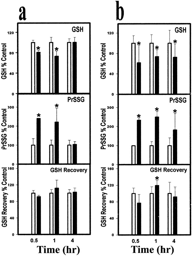
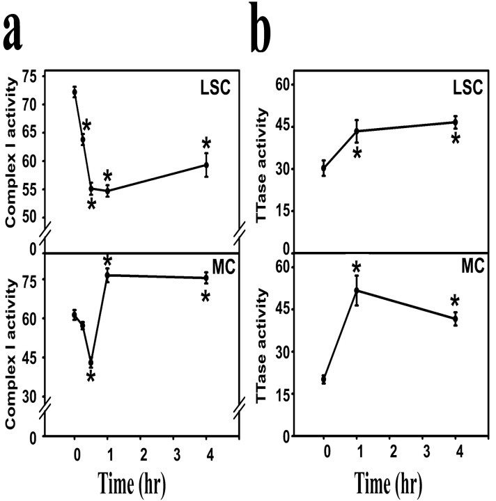
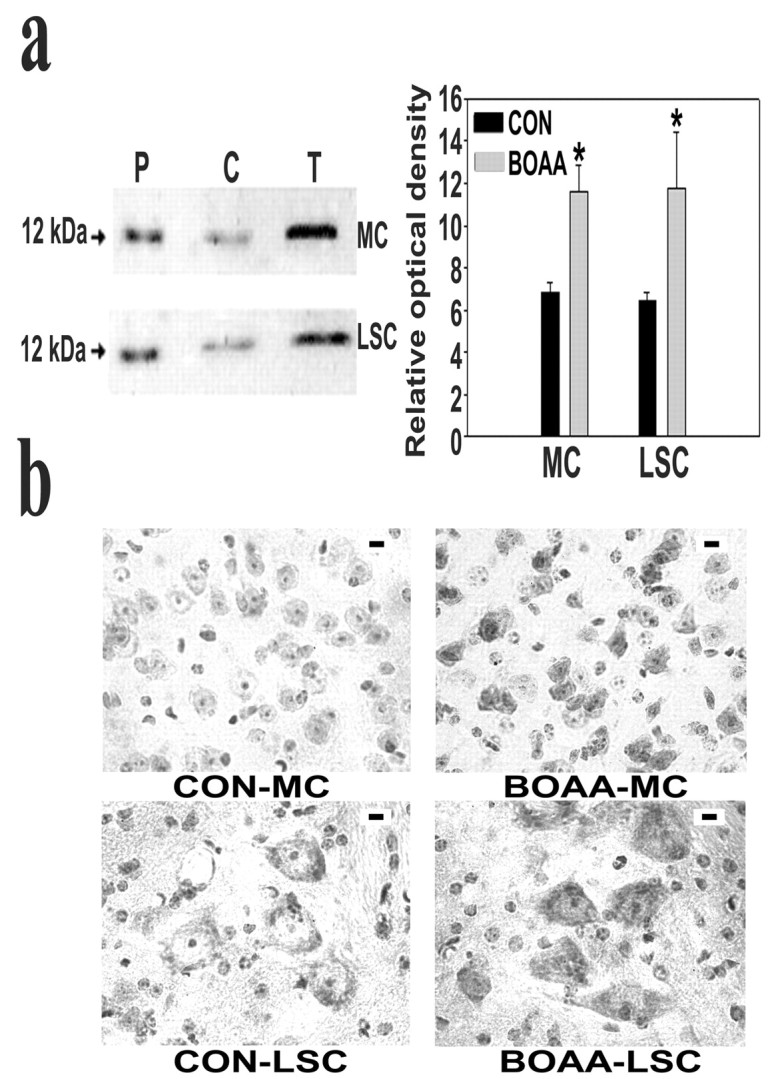
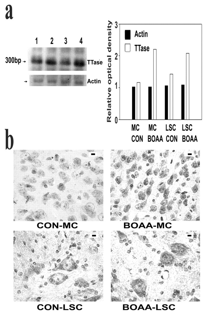
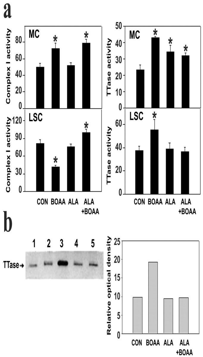
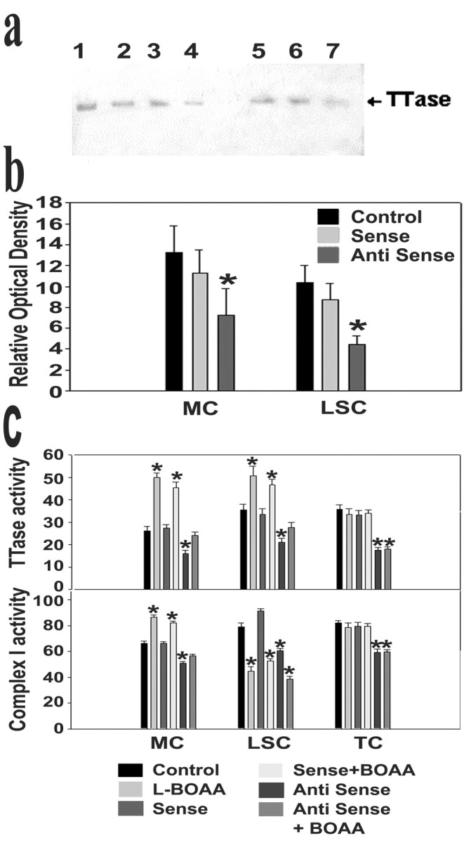
Mitochondrial dysfunction involving electron transport components is implicated in the pathogenesis of several neurodegenerative disorders and is a critical event in excitotoxicity. Excitatory amino acid L-beta-N-oxalylamino-L-alanine (L-BOAA), causes progressive corticospinal neurodegeneration in humans. In mice, L-BOAA triggers glutathione loss and protein thiol oxidation that disrupts mitochondrial complex I selectively in motor cortex and lumbosacral cord, the regions affected in humans. We examined the factors regulating postinjury recovery of complex I in CNS regions after a single dose of L-BOAA. The expression of thioltransferase (glutaredoxin), a protein disulfide oxidoreductase regulated through AP1 transcription factor was upregulated within 30 min of L-BOAA administration, providing the first evidence for functional regulation of thioltransferase during restoration of mitochondrial function. Regeneration of complex I activity in motor cortex was concurrent with increase in thioltransferase protein and activity, 1 hr after the excitotoxic insult. Pretreatment with alpha-lipoic acid, a thiol delivery agent that protects motor neurons from L-BOAA-mediated toxicity prevented the upregulation of thioltransferase and AP1 activation, presumably by maintaining thiol homeostasis. Downregulation of thioltransferase using antisense oligonucleotides prevented the recovery of complex I in motor cortex and exacerbated the mitochondrial dysfunction in lumbosacral cord, providing support for the critical role for thioltransferase in maintenance of mitochondrial function in the CNS.


Figures

Fig. 1.
Fig. 1.
GSH and PrSSG levels in motor cortex (a) and lumbosacral cord (b) at various periods after a single dose ofl-BOAA. l-BOAA (10 mg/kg body weight, s.c.) was administered to mice, and animals were killed after 0.5, 1, and 4 hr (black bars). Control animals received vehicle alone (white bars). GSH and PrSSG levels were estimated and are depicted as a percentage of corresponding controls. Total GSH recovered (sum of GSH equivalents recovered as GSH and as PrSSG) is also depicted. Values are mean ± SD (n = 3 animals). GSH levels in motor cortex and lumbosacral cord from control animals were 22.51 ± 0.823 and 21.41 ± 2.311 nmol/mg protein (equivalent to 1.54 ± 0.017 and 1.38 ± 0.168 μmol of GSH/gm tissue), respectively. PrSSG levels in motor cortex and lumbosacral cord of control animals were 1.71 ± 0.076 and 3.07 ± 0.135 nmol of GSH equivalents/mg protein (equivalent to 0.189 ± 0.083 and 0.203 ± 0.048 μmol of GSH equivalents/gm tissue), respectively. Asterisks indicate values significantly different from corresponding controls (p < 0.05).
Fig. 2.
Fig. 2.
Complex I (a) and thioltransferase (b) activities in the motor cortex (MC) and lumbosacral cord (LSC) at various periods after a single dose of l-BOAA. Mice were administered l-BOAA (10 mg/kg body weight, s.c.) and killed at indicated times. The control values were similar at all time points examined and are indicated as the zero hour value. Values are mean ± SEM (n = 6 animals). Complex I activity is expressed as nanomoles of NADH oxidized per minute per milligram of protein, and thioltransferase activity is expressed as nanomoles of NADPH oxidized per minute per milligram of protein.Asterisks indicate values significantly different from corresponding controls (p < 0.05).
Fig. 3.
Fig. 3.
Expression of thioltransferase protein in mouse CNS after l-BOAA treatment. Mice were killed 1 hr afterl-BOAA administration. a, A representative blot from motor cortex (MC) and lumbosacral cord (LSC) from control (C) andl-BOAA treated (T) animals subjected to immunoblot analysis using antiserum to thioltransferase (lanes contained 10 μg of protein). A sample of pure thioltransferase (P, 10 ng) was also loaded on the same gel. Densitometric analysis of the immunoblots representing the relative intensity of the immunoreactive bands from control (black bars) and l-BOAA-treated animals (gray bars) are represented. Values are mean ± SD (n = 3 animals). Asterisksindicate values significantly different from corresponding control (p < 0.05). b,Immunohistochemical localization of thioltransferase in motor cortex and lumbar cord revealed the increased levels of thioltransferase in the neurons in layer 3 of motor cortex (BOAA-MC) and the anterior horn cells of the lumbar cord (BOAA-LSC) as compared with untreated animals (CON-MC andCON-LSC) 1 hr after a single dose of l-BOAA. Scale bar, 25 μm.
Fig. 4.
Fig. 4.
Expression of thioltransferase mRNA in mouse CNS after l-BOAA treatment. Mice were administeredl-BOAA and killed 0.5 hr after l-BOAA.a, A representative Northern blot of the total RNA from motor cortex (lanes 1, 2; 10 μg) and lumbosacral cord (lanes 3,4; 10 μg) from control (lanes 1, 3) and treated (lanes 2, 4) animals, respectively, subjected to Northern blot analysis using cRNA to brain thioltransferase (TTase). The blots were also hybridized with β-actin cRNA for normalization. Densitometric analyses of the Northern blots representing the relative intensity of the hybridized bands from β-actin (black bars) and thioltransferase (white bars) are represented. Values are means of two individual experiments using pooled brain regions from three animals. b,In situ hybridization of thioltransferase mRNA in motor cortex and lumbar cord revealed increased levels of thioltransferase in the neurons in layer 3 of motor cortex (BOAA-MC) and the anterior horn cells of the lumbar cord (BOAA-LSC) as compared with untreated animals (CON-MC and CON-LSC) 0.5 hr afterl-BOAA. Scale bar, 25 μm.
Fig. 5.
Fig. 5.
Effect of α-lipoic acid pretreatment onl-BOAA-mediated inhibition of complex I activity and upregulation thioltransferase in mouse CNS. Animals were pretreated with α-lipoic acid (20 mg/kg body weight, s.c.) 1 hr before administration of l-BOAA (10 mg/kg body weight, s.c.) and killed 1 hr after the l-BOAA dose. a,Activities of complex I and thioltransferase were measured in the motor cortex and lumbosacral cord as described under Figure 1. Values are mean ± SD (n = 6 animals, andasterisks indicate values significantly different from vehicle-treated controls) (p < 0.05).b, A representative blot from motor cortex (10 μg) of animals treated with vehicle (lane 2),l-BOAA (lane 3), α-lipoic acid (lane 4), and α-lipoic acid andl-BOAA (lane 5) subjected to immunoblot analysis using antiserum to thioltransferase. A sample of pure thioltransferase (lane 1, 10 ng) was also loaded on the same gel. Densitometric analyses of immunoblots representing relative intensity of immunoreactive bands are represented. Values are mean of two individual experiments.
Fig. 6.
Fig. 6.
Activation of transcription factors AP1 (a), ARE (b), and NF-κB (c) after administration of l-BOAA to mice. Mice were administered l-BOAA and killed 15 min afterl-BOAA. Electrophoretic mobility shift assays were performed using nuclear extracts (5 μg of protein) from motor cortex (MC) and lumbar sacral cord (LSC) of mice treated with vehicle (C), l-BOAA (B), and α-lipoic acid and l-BOAA (A). Densitometric analyses of the shifted bands representing the relative intensity of binding of transcription factors are represented. Values are mean of three individual experiments.d, Antibody to phosphorylated cJun super shifts the mobility of the transcription factors AP1 after administration ofl-BOAA to mice. The nuclear extracts from motor cortex (MC; lanes 1–3) and lumbar sacral cord (LSC; lanes 4–6) of mice treated with vehicle (lanes 1, 4) and l-BOAA (lanes 2, 3,5, 6) were preincubated with antibody to phosphorylated c Jun before performing electrophoretic mobility shift assay as described above.
Fig. 7.
Fig. 7.
Effect of downregulation of thioltransferase protein using antisense oligonucleotides on l-BOAA-mediated mitochondrial dysfunction. Mice were injected intrathecally with sense and antisense oligonucleotides and killed 12 hr after the last injection. a, Immunoblot analysis of thioltransferase protein from motor cortex (lanes 2–4) and lumbosacral cord (lanes 5–7) from vehicle (lanes 2, 5), sense oligonucleotide-treated (lanes 3, 6), and antisense oligonucleotide-treated (lanes 4, 7) immunostained with antiserum to RBC thioltransferase (lanes contained 10 μg of protein). Lane 1 contained 10 ng of pure thioltransferase. b Represents the densitometric analysis of the immunoblots from three animals. Values are mean ± SD (n = 3 animals). c,Thioltransferase and complex I activities in motor cortex (MC), lumbosacral cord (LSC), and thoracic cord (TC) of animals treated with sense or antisense oligonucleotides (as described above) followed byl-BOAA administration and killed after 1 hr. Values are mean ± SD (n = 5–6) animals.Asterisks indicate values significantly different from vehicle-treated control (p < 0.05).

Similar articles

Cited by

References

    1. Akerboom TP, Sies H. Assay of glutathione, glutathione disulfide and glutathione mixed disulfides in biological samples. Methods Enzymol. 1981;77:373–382. - PubMed
    1. Annepu J, Ravindranath V. 1-Methyl-4-phenyl-1, 2, 3, 6 tetrahydropyridine induced complex I inhibition is reversed by disulfide reductant, dithiothreitol in mouse brain. Neurosci Lett. 2000;289:209–212. - PubMed
    1. Balijepalli S, Tirumalai PS, Swamy KV, Boyd MR, Mieyal JJ, Ravindranath V. Rat brain thioltransferase: regional distribution, immunological characterization and localization by fluorescent in situ hybridization. J Neurochem. 1999;72:1170–1178. - PubMed
    1. Balijepalli S, Boyd MR, Ravindranath V. Human brain thioltransferase: constitutive expression and localization by fluorescent in situ hybridization. Mol Brain Res. 2000;85:123–132. - PubMed
    1. Bradford MM. A rapid and sensitive method for the quantitation of microgram quantities of protein utilizing the principle of dye-binding. Anal Biochem. 1976;72:248–254. - PubMed

Publication types

MeSH terms

LinkOut - more resources