. 2002 Oct 1;22(19):8438-46.
doi: 10.1523/JNEUROSCI.22-19-08438.2002.

Structure/function analysis of Ca2+ binding to the C2A domain of synaptotagmin 1

Affiliations

Structure/function analysis of Ca2+ binding to the C2A domain of synaptotagmin 1

Rafael Fernández-Chacón et al. J Neurosci. .

Abstract

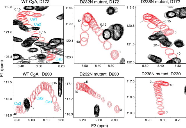
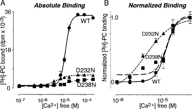
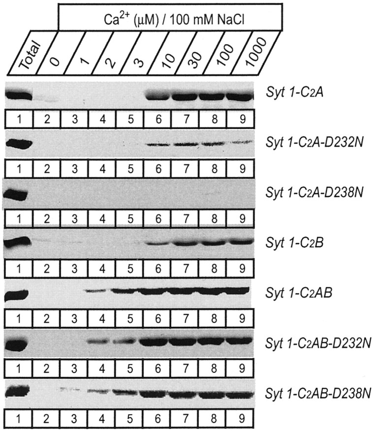
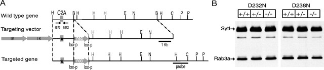
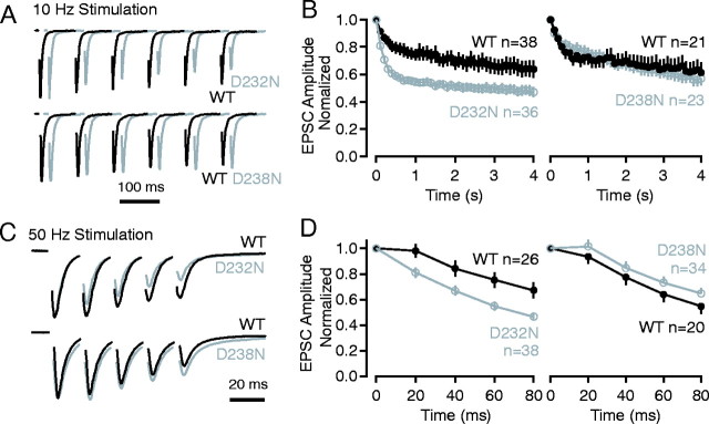
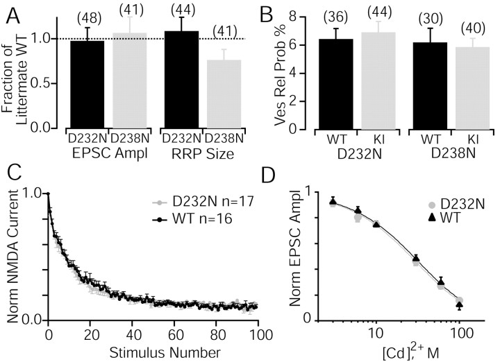
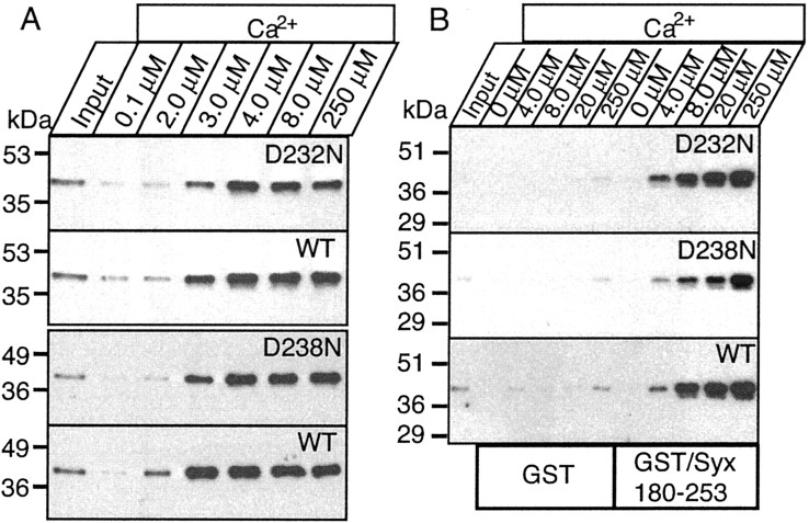
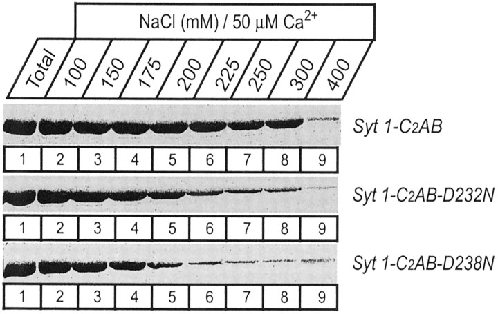
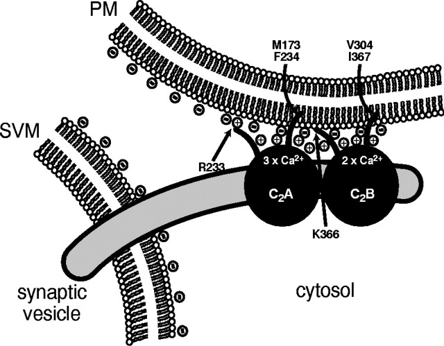
Synaptotagmin 1, a Ca2+ sensor for fast synaptic vesicle exocytosis, contains two C2 domains that form Ca2+-dependent complexes with phospholipids. To examine the functional importance of Ca2+ binding to the C2A domain of synaptotagmin 1, we studied two C2A domain mutations, D232N and D238N, using recombinant proteins and knock-in mice. Both mutations severely decreased intrinsic Ca2+ binding and Ca2+-dependent phospholipid binding by the isolated C2A domain. Both mutations, however, did not alter the apparent Ca2+ affinity of the double C2 domain fragment, although both decreased the tightness of the Ca2+/phospholipid/double C2 domain complex. When introduced into the endogenous synaptotagmin 1 gene in mice, the D232N and D238N mutations had no apparent effect on morbidity and mortality and caused no detectable alteration in the Ca2+-dependent properties of synaptotagmin 1. Electrophysiological recordings of cultured hippocampal neurons from knock-in mice revealed that neither mutation induced major changes in synaptic transmission. The D232N mutation, however, caused increased synaptic depression during repetitive stimulation, whereas the D238N mutation did not exhibit this phenotype. Our data indicate that Ca2+ binding to the C2A domain of synaptotagmin 1 may be important but not essential, consistent with the finding that the two C2 domains cooperate and may be partially redundant in Ca2+-dependent phospholipid binding. Moreover, although the apparent Ca2+ affinity of the synaptotagmin 1/phospholipid complex is critical, the tightness of the Ca2+/phospholipid complex is not. Our data also demonstrate that subtle changes in the biochemical properties of synaptotagmin 1 can result in significant alterations in synaptic responses.


Figures

Fig. 1.
Fig. 1.
Intrinsic Ca2+ binding to the wild-type (WT) and the D232N and D238N mutant C2A domains of synaptotagmin 1 monitored by NMR spectroscopy. Ca2+ titrations were examined by1H-15N HSQC spectra acquired with 150–165 μm of purified recombinant C2A domains.Panels illustrate the Ca2+-dependent shifts of selected cross-peaks corresponding to the D172 (top panels) and D230 (bottom panels) NH groups in all three C2A domains. The cross-peaks from these NH groups are shown in red, and other cross-peaks are shown in black. Numbers next to the resonances indicate the Ca2+ concentration (in millimolar) for that particular position of the cross-peak. Note the typical triphasic movement of the cross-peaks in the wild-type C2A domain, with the corresponding Ca2+-binding sites (Ca1,Ca2, and Ca3) indicated next to each phase. In the mutants, only biphasic movements with a different Ca2+ dependence are detectable. For quantitation of the various Ca2+-binding parameters, see Table1.
Fig. 2.
Fig. 2.
Apparent Ca2+-binding affinities of the wild-type (WT) and mutant C2A domains of synaptotagmin 1 measured by Ca2+-dependent GST pulldowns of radiolabeled liposomes. A, Ca2+ titrations of phospholipid binding with equal amounts of recombinant C2A domains (25 μg protein) and radiolabeled liposomes. Note that both mutations (D232N and D238N) inactivate >80% of Ca2+-dependent phospholipid binding, but that the remaining binding is still Ca2+ dependent.B, Same data as in B but normalized for maximal binding to illustrate the shift in apparent Ca2+ affinities of the D232N mutant. Note that because of the low signal-to-noise, in particular for the D238N mutant C2A domain, which exhibits almost no Ca2+-dependent phospholipid binding, the curve fits are rather inaccurate in single experiments as shown here but can be averaged from multiple experiments as summarized in Table 2.
Fig. 3.
Fig. 3.
Apparent Ca2+ affinities of wild-type and mutant single and double C2 domain fragments from synaptotagmin 1 measured by Ca2+-dependent binding to liposomes. The isolated wild-type and D232N and D238N mutant C2A domain, the wild-type C2B domain, and the wild-type and mutant double C2 domain fragments were analyzed. Liposomes composed of 25% PS/75% PC were incubated with the indicated C2 domains (present as soluble purified GST-fusion proteins) at the Ca2+ concentrations shown on top (clamped with Ca2+/EGTA buffers) and centrifuged, and bound proteins were analyzed by SDS-PAGE and Coomassie blue staining. Data shown are from a single representative experiment repeated multiple times.
Fig. 4.
Fig. 4.
Strategy for generating knock-in mice with D232N and D238N mutations in the synaptotagmin 1 C2A domain.A, Design of knock-in vectors for homologous recombination. Similar to previous experiments (Fernández-Chacón et al., 2001), a genomic clone containing a single exon from the murine synaptotagmin 1 gene (top) was used to generate targeting vectors in which D232N or D238N mutations were introduced into the exon (middle), with two copies of the thymidine kinase (TK) gene for negative selection and a neomycin resistance cassette (neo) for positive selection. The mutant exons were then introduced into the endogenous synaptotagmin 1 gene by homologous recombination, which also introduces the neomycin resistance gene cassette into the intron (bottom). Numbered arrows identify oligonucleotides (1872 and1873) used for genotyping. The position of the outside probe for detection of homologous recombination by Southern blotting is indicated on the right. The location of selected restriction sites are shown (H, HindIII;B, BglII; E,EcoRI; N, NheI;C, ClaI; P,PstI), and the scale is given on theright. The mutant mice produced by homologous recombination were then crossed with control mice obtained in a previous study (Fernández-Chacón et al., 2001) in which the neomycin gene was introduced into the intron without any mutations in the coding region, and all analyses were performed on littermate offspring from matings between double heterozygous mice carrying one mutant allele (either D232N or D238N) and one control allele with the neomycin cassette but without a mutation. B, Immunoblots of total brain homogenates (30 μg of protein) from wild-type (+/+), heterozygous (+/−), and homozygous (−/−) littermates stained with monoclonal antibodies specific for synaptotagmin 1 and rab3a.
Fig. 5.
Fig. 5.
Effect of the D232N and D238N mutations on short-term synaptic plasticity analyzed in cultured hippocampal neurons. A, Average synaptic currents (EPSCs) recorded from “microisland” cultures in response to 10 Hz stimulation. EPSCs are normalized to the first response; top traces show a comparison of D232N mutant neurons with wild-type (WT) neurons, and bottom traces show a comparison of D238N mutant neurons with wild-type neurons. Stimulus artifacts areblanked. B, Plot of the average EPSC amplitudes normalized to the first response recorded during 10 Hz stimulation. The comparison of D232N mutant with wild-type responses is shown on the left, and the comparison of D238N mutant with wild-type responses is shown on the right.C and D are same as A andB but with 50 Hz stimulation.
Fig. 6.
Fig. 6.
Analysis of the vesicle pools, release probabilities, and Ca2+ sensitivities in D232N and D238N mutant neurons in comparison with wild-type (WT) neurons. A, Quantitative analysis of evoked responses and the sizes of the readily releasable vesicle pools of excitatory neurons of D232N and D238N excitatory neurons. Mean amplitudes and pool sizes were normalized to the mean values from the littermate wild-type neurons. B, The vesicular release probability was then calculated as the percentage of action potential evoked vesicles compared with the readily releasable pool. Data shown are means ± SEMs from the number of cells shown in parentheses in the bars. C, Evaluation of synaptic release probability from D232N and wild-type neurons. The rate of use-dependent block of NMDA-EPSCs in the presence of the irreversible open channel blocker MK-801 (5 μm) was not different between D232N and wild-type neurons, indicating that release probability was unchanged by the mutation. D, Ca2+ dependence of evoked release. EPSCs were evoked in 12 mm external Ca2+ and 1 mm Mg2+. Presynaptic Ca2+ influx was varied by adding various concentrations of Cd2+ (3–100 μm) to the external medium (n = 7–35 per concentration).Solid lines are best fits to a logistic hill function to determine the IC50 value for Cd2+.
Fig. 7.
Fig. 7.
The D232N and D238N mutations do not cause a significant change in Ca2+-dependent phosphopholipid and syntaxin binding by native synaptotagmin 1. A, Soluble C2A/C2B domain fragments of native synaptotagmin 1 were obtained from wild-type and mutant littermate mice by partial trypsin digestion of brain membranes followed by centrifugation. The soluble C2A/C2B domain fragment released into the supernatant was used for binding experiments with liposomes at the indicated free Ca2+ concentrations. Input and bound proteins were then analyzed by immunoblotting using a monoclonal synaptotagmin 1 antibody. The two mutations were analyzed in independent experiments with their littermate wild-type controls, resulting in separate wild-type controls for each mutant. Numbers on theleft indicate positions of size markers.B, GST-pulldown experiments of the soluble C2A/C2B domain fragment of synaptotagmin 1 isolated as described above. Proteins were bound to GST and GST-syntaxin (residues 180–264) at the indicated concentrations of free Ca2+ as described in A, and bound proteins were visualized by immunoblotting.
Fig. 8.
Fig. 8.
Salt sensitivity of the Ca2+-dependent complex of the wild-type and mutant double C2 domain fragment from synaptotagmin 1. The double C2 domain fragments were bound to liposomes in the presence of 50 μm Ca2+ and the indicated concentrations of NaCl. Double C2 domain proteins attached to the liposomes after centrifugation were analyzed by SDS-PAGE and Coomassie staining. Note especially that the D238N mutation destabilized the Ca2+-dependent phospholipid complex.
Fig. 9.
Fig. 9.
Model of the Ca2+-dependent binding of synaptotagmin 1 C2 domains to phospholipid membranes. The two C2 domains of synaptotagmin 1 (which account for two-thirds of the total sequence) engage in similar but parallel interactions with phospholipid membranes that are fueled by three forces: positive charges supplied by bound Ca2+ ions that are sandwiched between negatively charged phospholipid head groups and C2 domain aspartate residues; positive charges supplied by arginine and lysine residues such as R233 and K366 at the top of the domain; and hydrophobic residues that insert at least partially into the bilayer, such as M173, F234, V305, and I367. In the absence of Ca2+, repulsion by the negatively charged aspartate residues on top of the C2 domains and the negatively charged phospholipid head groups prevents the positively charged and hydrophobic residues at the top of the domain to engage in interactions. Thus in addition to forming a bridge between the phospholipid head groups and the top loops of the C2 domains, Ca2+ ions also neutralize repulsive negative charges. The double C2domains exhibit an approximately threefold higher apparent Ca2+ affinity than the individual isolated C2 domains when the two independent C2 domains become linked physically.

Similar articles

Cited by

References

    1. Bai J, Earles CA, Lewis JL, Chapman ER. Membrane-embedded synaptotagmin penetrates cis or trans target membranes and clusters via a novel mechanism. J Biol Chem. 2000;275:25427–25435. - PubMed
    1. Bai J, Wang P, Chapman ER. C2A activates a cryptic Ca2+-triggered membrane penetration activity within the C2B domain of synaptotagmin 1. Proc Natl Acad Sci USA. 2002;99:1665–1670. - PMC - PubMed
    1. Bollmann JH, Sakmann B, Borst JG. Calcium sensitivity of glutamate release in a calyx-type terminal. Science. 2000;289:953–957. - PubMed
    1. Chapman ER, Davis AF. Direct interaction of a Ca2+-binding loop of synaptotagmin with lipid bilayers. J Biol Chem. 1998;273:13995–14001. - PubMed
    1. Chapman ER, Jahn R. Calcium-dependent interaction of the cytoplasmic region of synaptotagmin with membranes. Autonomous function of a single C2-homologous domain. J Biol Chem. 1994;269:5735–5741. - PubMed

Publication types

MeSH terms

LinkOut - more resources