. 2002 Oct 1;22(19):8458-65.
doi: 10.1523/JNEUROSCI.22-19-08458.2002.

Activation of Bcl-2-associated death protein and counter-response of Akt within cell populations during seizure-induced neuronal death

Affiliations

Activation of Bcl-2-associated death protein and counter-response of Akt within cell populations during seizure-induced neuronal death

David C Henshall et al. J Neurosci. .

Abstract

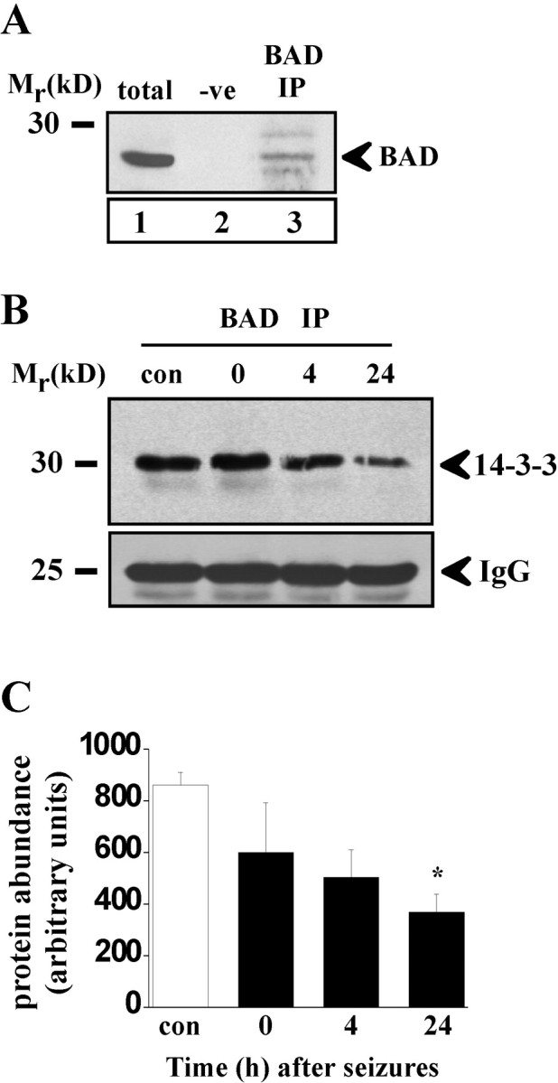
Bcl-2 family gene products are critical to the integration of cell death stimuli that target the mitochondrion. Proapoptotic BAD (Bcl-2-associated death protein) has been shown to dissociate from its sequestered site with the molecular chaperone protein 14-3-3 and displace proapoptotic BAX (Bcl-2-associated X protein) from antiapoptotic BCL-Xl. BAX subsequently translocates to the mitochondrion and induces cytochrome c release and caspase activation. Herein we report the response of the key members of this proposed pathway after seizures. Seizures evoked by microinjection of kainic acid into the amygdala of the rat induced unilateral CA3 pyramidal neuron death with features of apoptosis. In control hippocampus and cortex, BAD was found constitutively bound to 14-3-3, whereas BCL-Xl bound BAX. Within damaged hippocampus, seizures induced the dissociation of BAD from 14-3-3 and the subsequent dimerization of BAD with BCL-Xl as determined by immunoprecipitation and immunohistochemical colocalization. 14-3-3 was found to translocate to the nucleus of degenerating neurons, whereas BAX accumulated at mitochondrial membranes. In contrast, the primarily uninjured cortex exhibited increased phosphorylation of Akt (protein kinase B), which may phosphorylate and inhibit BAD, and no altered binding of BAD to BCL-Xl. Finally, administration of an inhibitor of phosphatidylinositol 3-kinase (LY294002), thought to be an upstream activator of Akt, exacerbated cortical apoptosis after seizures. These data suggest that seizures elicit divergent cell death and survival responses within neuronal populations and that the BAD cell death pathway may perform an instigator or reinforcement role in seizure-induced neuronal death.


Figures

Fig. 1.
Fig. 1.
BAD pathway and seizure-induced neuronal injury characteristics. A, Schematic depicting BAD cell death pathway, its binding interaction changes, and putative pharmacological manipulation. The pathway is thought to be engaged after the following: (1) calcium (Ca2+) entry, which activates the phosphatase calcineurin, which then (2) dephosphoryates BAD. Released BAD (3) then interacts with (4) the BCL-X:BAX complex and displaces BAX, resulting in (5) a BAD:BCL-X dimer. (6) BAX subsequently translocates to the mitochondrion, in which it triggers (7) cytochrome c release and formation of the apoptosome, which results in apoptosis. In turn, sequestration of BAD to 14-3-3 may be restored by phosphorylation downstream of (8) Akt. NMDAR, NMDA receptor; P, phosphate group; PP2B, protein phosphatase 2B/calcineurin; CASP, caspase;Akt, protein kinase B; PI3K, PI3-kinase;Cyt c, cytochrome c;APAF-1, apoptotic protease-activating factor 1.B, Histopathology of seizure model. a, Photomicrograph showing control ipsilateral hippocampus of the rat.Inset, High-power magnification of a single CA3 pyramidal neuron. b, Toluidine blue-stained CA3 field revealing early (4 hr) degenerative changes in some CA3 neurons.c, Photomicrograph 24 hr after seizures revealing marked loss of CA3 pyramidal neurons. Inset, Shrunken, condensed nuclei of two CA3 cells. d, Low-power field showing extensive TUNEL labeling (DNA fragmentation) throughout the degenerating CA3 subfield. Inset, View of a CA3 TUNEL-labeled cell exhibiting nuclear features of apoptosis. Scale bar: a, c, d, 200 μm; b, 15 μm.
Fig. 2.
Fig. 2.
Expression of BAD cell death pathway components after seizures. Representative Western blots (n = 2 per lane) showing expression of key components of BAD pathway within ipsilateral hippocampus. Seizures had no obvious effect on expression levels of any of the BAD pathway components. Protein-loading controls are shown for α-tubulin. Molecular weight markers are depicted to theleft. Immunoblots are representative of at least two independent experiments.
Fig. 3.
Fig. 3.
Dissociation of BAD from 14-3-3 after seizures.A, Representative Western blot (n = 2 per lane) to demonstrate effectiveness of BAD antibody to precipitate BAD. Lane 1, BAD in rat brain (total); lane2, negative control (−ve) in which the immunoprecipitation antibody was omitted; lane3, confirmation that the BAD antibody effectively precipitated BAD from rat brain (BAD IP). B, Representative Western blot showing decline of 14-3-3 binding to BAD after seizures (n = 4 per lane) within ipsilateral hippocampus. IgG bands are shown to confirm equality of antibody loading.C, Semiquantification of BAD interaction with 14-3-3 within ipsilateral hippocampus confirming decreased binding of BAD to 14-3-3 after seizures. Data are from two independent experiments. *p < 0.05 compared with control.
Fig. 4.
Fig. 4.
Immunohistochemical colocalization of BAD with 14-3-3 and their dissociation after seizures. A, Control expression of 14-3-3 was detected throughout cortex (CTX) and colocalized substantially with BAD (B) as evidenced by image overlay (C). Twenty-four hours after seizures, 14-3-3 expression (D) became almost exclusively nuclear within CA3 pyramidal neurons. In contrast, examination of a single BAD-labeled CA3 cell (E, F) revealed punctate staining that remained within the cytoplasm. Counterstaining of 14-3-3-labeled sections (G) with the DNA fragmentation marker TUNEL (H) revealed that nuclear 14-3-3 was exclusively within dying cells. Arrowheads indicate representative immunopositive cells. Scale bars: A–C,D, 15 μm; E, F, 4 μm;G–I, 18 μm.
Fig. 5.
Fig. 5.
Increased binding of BAD to BCL-Xl after seizures.A, Representative Western blot (n = 4 per lane) showing increased binding of BCL-Xl to BAD immunoprecipitates (BAD IP) 4 hr after seizures within ipsilateral hippocampus compared with control (con). Whole brain lysate (total) and omission of the immunoprecipitated antibody (−ve) are shown to confirm reaction specificity. B, Semiquantification of increased BAD interaction with BCL-Xl after seizures from two independent experiments. *p < 0.05 compared with control. C, Immunohistochemical colocalization of BAD (green) and BCL-Xl (red) 24 hr after seizures within CA3 as revealed byyelloworange coloring inmergepanel. Insets show corresponding control expression of each protein. Arrowheadsindicate representative immunopositive cells. Scale bar, 15 μm.
Fig. 6.
Fig. 6.
BAX accumulation at mitochondria after seizures.A, Immunohistochemical detection of BAX (a; red) and the mitochondrial marker COXIV (green) within control brain CA3 subfield in which no colocalization is detected. Twenty-four hours after seizures, BAX (b) appeared as a punctate stain within affected CA3 cells and exhibited marked colocalization with COXIV (c) as revealed byyellow–gold puncta in d, themergepanel. Inset ind shows colocalization at 4 hr in which overlap is detectable but not as extensive. Arrowheads indicate representative immunopositive cells. Scale bar, 15 μm.B, Representative electron micrographs showing (a, low-power field; b, high-power field) BAX accumulation at mitochondrial (mt) membranes 2 hr after seizures as labeled by 15 nm immunogold particles. Note clustering of BAX at outer surface of mitochondria.
Fig. 7.
Fig. 7.
Effects of FK506 on cell survival.A, Graph showing quantification of hippocampal CA3 TUNEL labeling in control (con) and seizure (seiz) animals injected with vehicle or FK506 (n = 6 per group). Seizures induced significant TUNEL labeling within CA3 cells 72 hr after diazepam compared with nonseizure controls. FK506 (seiz + FK) significantly reduced numbers of TUNEL-positive cells compared with vehicle-treated seizure animals. B, Seizures induced a significant reduction in numbers of CA3 neurons after seizures, and this was partly blocked by FK506.
Fig. 8.
Fig. 8.
Akt activation in surviving brain regions after seizures. A, Representative Western blots (n = 2 per lane) showing Akt expression and phosphoAkt Thr308 and Ser473levels in ipsilateral hippocampus after seizures. No activation of Akt was detected after seizures. B, Representative Western blots (n = 2 per lane) showing Akt expression and rapid phosphorylation at both consensus sites within cortex after seizures. C, Immunohistochemical detection of phosphoAkt Ser473 in rat brain revealed low to undetectable levels of phosphoAkt473 within control cortex (a). b, Twenty-four hours after seizures, phosphoAkt473 was upregulated within many cells within cortex (CTX). Arrowheadsindicate representative immunopositive cells. Scale bar (ina): 20 μm. D, Quantification of TUNEL counts 72 hr after seizures in control animals (con) or seizure rats infused with vehicle (seiz) or the PI3-kinase inhibitor LY294002 (seiz + LY) (n = 5 per group). a, Seizures induced significant hippocampal CA3 TUNEL labeling compared with nonseizure controls in both groups, but there was no significant difference between vehicle and LY294002 groups. In contrast, LY294002 (b) significantly increased numbers of TUNEL counts within cortex compared with both nonseizure controls and vehicle-treated seizure animals.

Similar articles

Cited by

References

    1. Adams JM, Cory S. The Bcl-2 protein family: arbiters of cell survival. Science. 1998;281:1322–1326. - PubMed
    1. Bengzon J, Kokaia Z, Elmer E, Nanobashvili A, Kokaia M, Lindvall O. Apoptosis and proliferation of dentate gyrus neurons after single and intermittent limbic seizures. Proc Natl Acad Sci USA. 1997;94:10432–10437. - PMC - PubMed
    1. Bratton SB, Walker G, Srinivasula SM, Sun XM, Butterworth M, Alnemri ES, Cohen GM. Recruitment, activation and retention of caspases-9 and -3 by Apaf-1 apoptosome and associated XIAP complexes. EMBO J. 2001;20:998–1009. - PMC - PubMed
    1. Burgering BM, Coffer PJ. Protein kinase B (c-Akt) in phosphatidylinositol-3-OH kinase signal transduction. Nature. 1995;376:599–602. - PubMed
    1. Butcher SP, Henshall DC, Teramura Y, Iwasaki K, Sharkey J. Neuroprotective actions of FK506 in experimental stroke: in vivo evidence against an antiexcitotoxic mechanism. J Neurosci. 1997;17:6939–6946. - PMC - PubMed

Publication types

MeSH terms