. 2002 Oct 1;22(19):8532-40.
doi: 10.1523/JNEUROSCI.22-19-08532.2002.

Caspase activation in hair cells of the mouse utricle exposed to neomycin

Affiliations

Caspase activation in hair cells of the mouse utricle exposed to neomycin

Lisa L Cunningham et al. J Neurosci. .

Abstract

Aminoglycoside exposure results in the apoptotic destruction of auditory and vestibular hair cells. This ototoxic hair cell death is prevented by broad-spectrum caspase inhibition. We have used in situ substrate detection, immunohistochemistry, and specific caspase inhibitors to determine which caspases are activated in the hair cells of the adult mouse utricle in response to neomycin exposure in vitro. In addition, we have examined the hierarchy of caspase activation. Our data indicate that both upstream caspase-8 and upstream caspase-9, as well as downstream caspase-3 are activated in hair cells exposed to neomycin. The inhibition of caspase-9-like activity provided significant protection of hair cells exposed to neomycin, whereas the inhibition of caspase-8-like activity was not effective in preventing neomycin-induced hair cell death. In addition, caspase-9 inhibition prevented the activation of downstream caspase-3, whereas the inhibition of caspase-8 did not. These data indicate that caspase-9 is the primary upstream caspase mediating neomycin-induced hair cell death in this preparation.


Figures
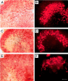
Fig. 1.
Fig. 1.
Cultured utricles double-labeled for calmodulin and calbindin. The utricles were cultured for 24 hr without neomycin (A, B), with 1 mm neomycin (C, D), or with 5 mm neomycin (E, F). After the culture period the utricles were fixed and double-labeled using antibodies directed against calmodulin (A, C, E), which labels all hair cells in the utricle, and calbindin (B, D, F), which labels the type I hair cells in the striolar region. Exposure to 1 mm neomycin (C, D) resulted in significant loss of striolar hair cells compared with control cultures. At a dose of 5 mmneomycin (E, F) the majority of hair cells are missing in both the extrastriolar and striolar regions. Scale bar, 50 μm.
Fig. 2.
Fig. 2.
Relationship between neomycin concentration and hair cell density. The utricles were cultured for 24 hr in various concentrations of neomycin and then double-labeled for calmodulin and calbindin. The hair cells in each utricle were counted and averaged from four striolar and four extrastriolar regions of 900 μm2 each. The hair cell density is expressed as a percentage of the control value (cultured without neomycin). The hair cell density decreased systematically as the neomycin concentration increased. For comparison, the unconnected filled symbols show hair cell densities for utricles that were not cultured. Mean hair cell densities ± SEM, n = 5–10 utricles per group.
Fig. 3.
Fig. 3.
Protection of hair cells by broad-spectrum caspase inhibition. The utricles were cultured for 24 hr in control media (n = 5), 1 mm neomycin (n = 8), or 1 mm neomycin plus 100 μm z-VAD-fmk (n = 5), a broad-spectrum caspase inhibitor. The utricles were double-labeled for calmodulin and calbindin, and the hair cells were counted. z-VAD-fmk provided significant protection of hair cells in both the striolar and extrastriolar regions compared with utricles cultured in neomycin alone (*p < 0.05, one-way ANOVA). Error bars indicate ±SEM.
Fig. 4.
Fig. 4.
Caspase-3 substrate activation. The utricles were cultured for 12 hr in control media (A) or 1 mm neomycin (B). Thegreen fluorescent caspase-3 substrate fam-DEVD-fmk was added for the final hour in culture. After the culture period the utricles were fixed and stereocilia were labeled using rhodamine phalloidin. Shown are brightest point projections ofz-series confocal micrographs that were limited to a depth of 5 μm in the z-axis. A, Very little or no activation of caspase-3 is seen in control utricles.B, Robust caspase-3 activation is seen in utricles cultured in the presence of neomycin. Scale bar, 20 μm.
Fig. 5.
Fig. 5.
Caspase-9 activation. The utricles were cultured for 12 hr in control media (A, C) or 1 mmneomycin (B, D). Caspase-9 activation (green) was detected using the fluorescent caspase-9 substrate fam-LEHD-fmk (A, B) and immunohistochemistry (C, D). Stereocilia were labeled using rhodamine phalloidin. Shown are brightest point projections ofz-series confocal micrographs that were limited to a depth of 5 μm in the z-axis. A, Little activation of caspase-9 is seen in control utricles using the substrate detection assay. B, Robust caspase-9 activation is seen in utricles cultured in the presence of neomycin. C, D,Immunochemistry for active caspase-9 shows very little activation in control utricles (C) and marked activation (arrows) in utricles cultured in neomycin (D). Scale bars: A, B, 10 μm;C, D, 5 μm.
Fig. 6.
Fig. 6.
Caspase-8 activation. The utricles were cultured for 12 hr in control media (A, C) or 1 mmneomycin (B, D). After the culture period the utricles were fixed, and stereocilia were labeled using rhodamine phalloidin. Shown are brightest point projections of z-series confocal micrographs that were limited to a depth of 5 μm in thez-axis. A, B, Thegreen fluorescent caspase-8 substrate fam-LETD-fmk was added for the final hour in culture. Limited activation of caspase-8 is seen in the hair cells of control utricles (A). Caspase-8 activation is increased in the utricles cultured in the presence of neomycin (B). C, D, Immunochemistry using an antibody specific for the activated form of caspase-8. Very little active caspase-8 immunoreactivity is detectable in control utricles (C). Active caspase-8 immunoreactivity (arrows) is present in the hair cells of utricles cultured in neomycin (D). Scale bars: A, B, 20 μm; C, D, 5 μm.
Fig. 7.
Fig. 7.
Quantification of active caspase-8 substrate detection data. Hair cells showing caspase-8 activation by the substrate detection method were counted in utricles cultured with and without 1 mm neomycin. Significantly more hair cells contained activated caspase-8 in utricles cultured in neomycin compared with control utricles (*p < 0.01; n = 5 utricles per condition). Error bars indicate means ± SD.
Fig. 8.
Fig. 8.
Inhibition of caspase-9 and caspase-8. The utricles were cultured for 24 hr in control media, neomycin alone, or neomycin plus a peptide inhibitor of either caspase-9-like or caspase-8-like activity. A, The utricles were cultured in control media (n = 6), 1 mmneomycin (n = 8), or 1 mm neomycin plus z-LEHD-fmk, a peptide inhibitor of caspase-9, at concentrations of 100 μm (n = 7), 50 μm(n = 4), or 25 μm(n = 5). Hair cell densities were calculated for the striolar and extrastriolar regions. 100 μm z-LEHD-fmk provided significant protection of hair cells in both the striolar and extrastriolar regions (*p < 0.05 for the extrastriolar region, *p < 0.001 for the striolar region, ANOVA). Hair cells of the striolar region (but not the extrastriolar region) were significantly protected by 50 μm z-LEHD-fmk compared with control utricles (*p < 0.05 for the striolar region, ANOVA). No significant protection of hair cells was seen at 25 μmz-LEHD-fmk. B, The utricles were cultured in control media (n = 6), 1 mm neomycin (n = 6), or 1 mm neomycin plus z-IETD-fmk, a peptide inhibitor of caspase-8-like activity, at either 100 μm (n = 5) or 50 μm(n = 5) inhibitor. Caspase-8 inhibition did not result in significant protection of hair cells of either the striolar or extrastriolar region. Note: Although it appears that the means of the 1 mm condition differ betweenA and B, they are within the normal variability and were not statistically different from one another (p > 0.05 for both extrastriolar and striolar regions). Error bars indicate means ± SEM.
Fig. 9.
Fig. 9.
Inhibition of caspase-8 or caspase-9 with substrate detection of activated caspase-3. The utricles were cultured for 12 hr in control media (A), in 1 mm neomycin alone (B), in 1 mm neomycin plus the caspase-8 inhibitor z-IETD-fmk (100 μm) (C), or in 1 mmneomycin plus the caspase-9 inhibitor z-LEHD-fmk (100 μm) (D). In all panels the greenfluorescent caspase-3 substrate fam-DEVD-fmk was used to examine caspase-3 activation. Hair cell stereocilia were labeled using rhodamine phalloidin. Shown are the brightest point projections ofz-series confocal micrographs that were limited to a depth of 5 μm in the z-axis. A, In the absence of neomycin there is very little activation of caspase-3 in hair cells. B, In the presence of neomycin alone there is robust activation of caspase-3 in hair cells. C, In the presence of neomycin and z-IETD-fmk (caspase-8 inhibitor), there is robust activation of caspase-3 in hair cells. D, In the presence of neomycin and z-LEHD-fmk (caspase-9 inhibitor) there is very little activation of caspase-3 in hair cells. Scale bar, 20 μm.
Fig. 10.
Fig. 10.
Quantification of caspase-3-positive hair cells in utricles cultured in the presence of inhibitors of caspase-8 or caspase-9. The utricles were cultured as described in Figure 9. Caspase-3-positive hair cells were counted for utricles cultured in control media, neomycin alone, or neomycin plus peptide inhibitors of either caspase-8-like activity (z-IETD-fmk) or caspase-9-like activity (z-LEHD-fmk). The inhibition of caspase-9 resulted in a significant reduction in the number of active caspase-3-positive hair cells compared with utricles cultured in neomycin alone (*p < 0.01; ANOVA; n = 7 utricles). Inhibition of caspase-8 did not result in any significant change in the number of caspase-3-positive hair cells compared with incubation in neomycin alone (p = 0.7; ANOVA; n = 5 utricles). Error bars indicate means ± SD.

References

    1. Atlante A, Calissano P, Bobba A, Azzariti A, Marra E, Passarella S. Cytochrome c is released from mitochondria in a reactive oxygen species (ROS)-dependent fashion and can operate as a ROS scavenger and as a respiratory substrate in cerebellar neurons undergoing excitotoxic death. J Biol Chem. 2000;275:37159–37166. - PubMed
    1. Bagger-Sjoback D, Wersall J. Gentamicin-induced mitochondrial damage in inner ear sensory cells of the lizard Calotes versicolor. Acta Otolaryngol. 1978;86:35–51. - PubMed
    1. Bedner E, Smolewski P, Amstad P, Darzynkiewicz Z. Activation of caspases measured in situ by binding of fluorochrome-labeled inhibitors of caspases (FLICA): correlation with DNA fragmentation. Exp Cell Res. 2000;259:308–313. - PubMed
    1. Bhave SA, Oesterle EC, Coltrera MD. Macrophage and microglia-like cells in the avian inner ear. J Comp Neurol. 1998;398:241–256. - PubMed
    1. Bossy-Wetzel E, Green DR. Apoptosis: checkpoint at the mitochondrial frontier. Mutat Res. 1999;434:243–251. - PubMed

Publication types

MeSH terms