. 2002 Oct 1;22(19):8541-52.
doi: 10.1523/JNEUROSCI.22-19-08541.2002.

Refinement of thalamocortical arbors and emergence of barrel domains in the primary somatosensory cortex: a study of normal and monoamine oxidase a knock-out mice

Affiliations

Refinement of thalamocortical arbors and emergence of barrel domains in the primary somatosensory cortex: a study of normal and monoamine oxidase a knock-out mice

Alexandra Rebsam et al. J Neurosci. .

Abstract

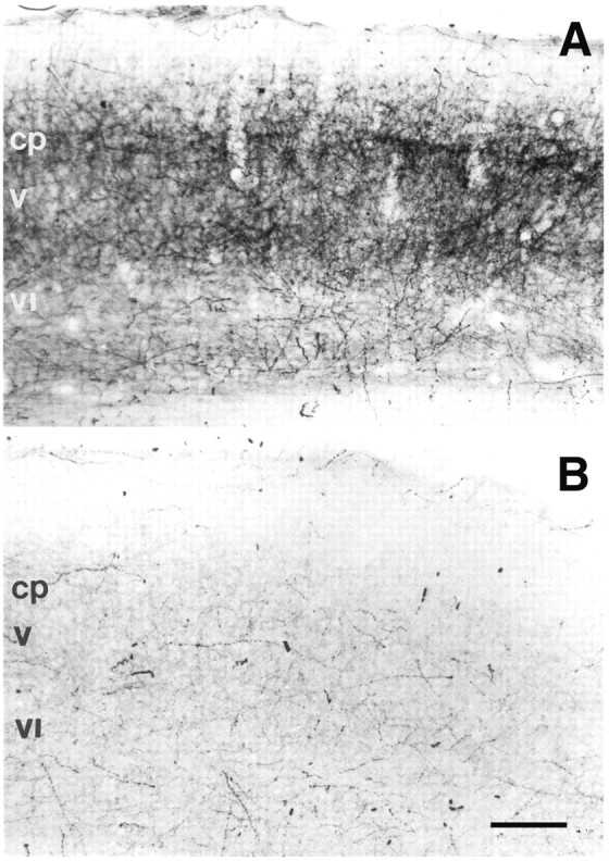
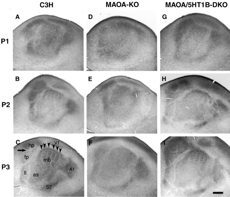
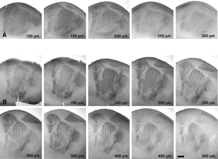
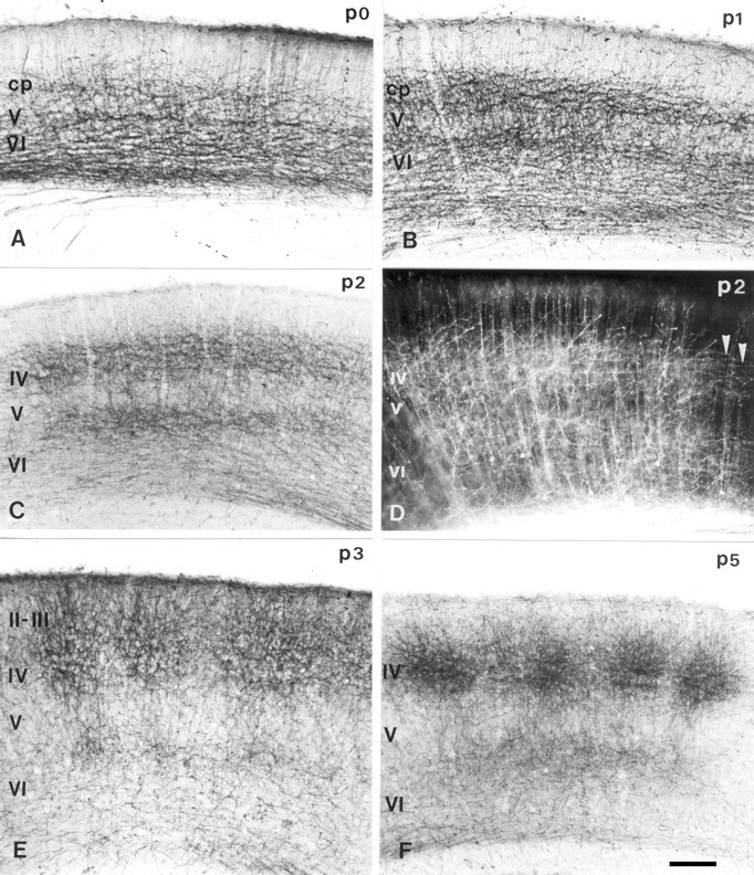
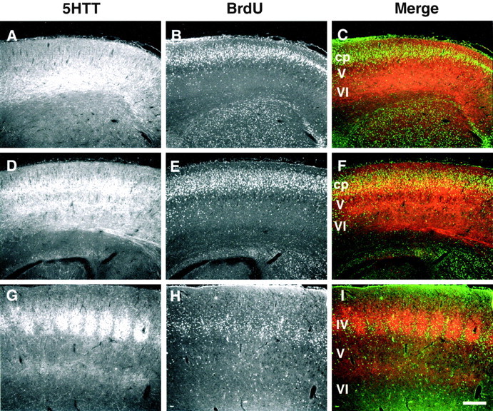
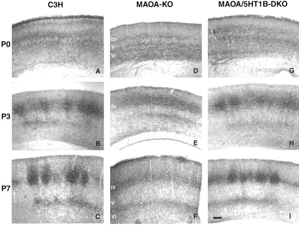
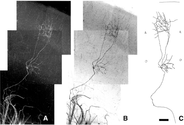
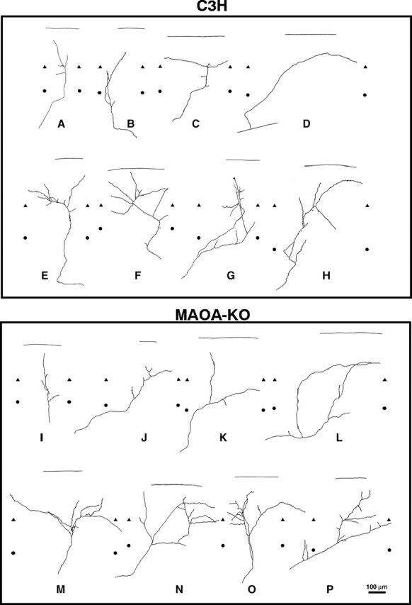
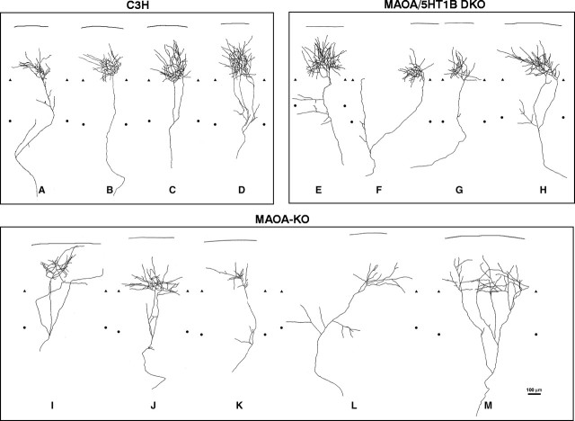
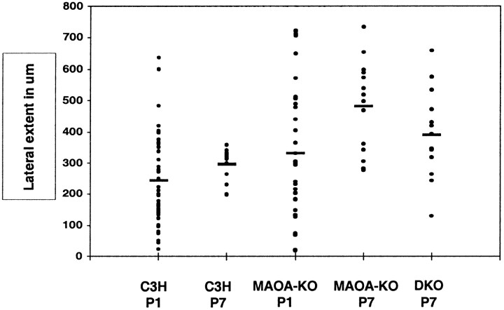
In the rodent primary somatosensory cortex, the thalamocortical axons (TCAs) are organized into clusters that correspond to functional units in the periphery. Around these axons, neurons in layer IV aggregate as barrels. To understand how this organization emerges, we analyzed TCA development in mice that do not form barrels, the monoamine oxidase A knock-out (MAOA-KO), and in MAOA/5-HT(1B) receptor double-KO mice, which have a restored barrel field. We show that TCAs already attain cortical layer IV on the day of birth. They are uniformly distributed in this layer from postnatal day 0 (P0) to P2 and secondarily coalesce into barrel domains in layer IV, over a 3 d period (P3-P5), with no prepatterning in the deeper layers. In MAOA-KO mice, the uniform distribution of the TC projection is maintained, and no axon clusters emerge. Individual TCA arbors were traced after carbocyanine injections. At P1, TCAs were poorly branched and covered variable tangential widths, encompassing one to two prospective barrels. At P7 the number of TCA branches increased 10-fold in layer IV and became restricted to one barrel. In MAOA-KO mice, there was a 50% reduction of the TCA terminal branches in layer IV, with a 40% increase in their tangential extent. These defects were corrected in the MAOA/5-HT(1B) double knock-out mice, indicating an effect of the presynaptic 5-HT(1B) receptor on axon branching. Our results indicate that the barrel-deficient phenotype of MAOA-KO mice results from an altered refinement of the TCA arbors in their target layer IV, involving branch elaboration and collateral retraction during early postnatal life.


Figures

Fig. 1.
Fig. 1.
5-HT immunostaining preferentially labels thalamocortical axons at P1. During development, 5-HT is present in fibers arising from the raphe as well as in TCAs, which express the 5-HT transporter (Lebrand et al., 1996). A, At P1, the 5-HT labeling is distributed as two dense bands, one in the cortical plate (cp) and another at the border between layers V and VI, with a pattern similar to that of anterogradely labeled TCAs shown in Figure 4D. B, To abolish 5-HT staining in the TCAs, we treated acutely mice with fluoxetine, an inhibitor of 5-HT uptake. Raphe fibers, which synthesize 5-HT, are not affected by this treatment. They are much less abundant than the TCAs and are sparsely distributed in the cortical plate (cp) and layers V to VI and I. Scale bar, 100 μm.
Fig. 2.
Fig. 2.
Progressive individualization of tangential domains in S1. In the three genotypes, WT (A–C), MAOA-KO (D–F), and MAOA/5-HT1B-DKO (G–I) mouse pups from the same litters were analyzed at P1, P2, and P3. 5-HTT immunostaining was performed on serial tangential sections of flattened hemispheres. All pictures are similarly oriented; rostral is to the left, and dorsal is up. 5-HTT labels all of the TC projection of the sensory cortices, including the somatosensory (S1, S2), visual (V1), and auditory (A1) cortices. The main divisions in S1 are shown in C: the mystacial vibrissa subfield (mb), anterior snout (as), lower lip (ll), hindpaw (hp), and forepaw (fp). A–C, In the C3H WT mice, the emergence of tangential domains is sequential; in the mystacial vibrissae field, a diffuse pattern of staining is observed at P1, barrel rows emerge at P2, and individual barrels emerge at P3 (arrowheads show the limits between the barrel rows). Note that the barrels are first delineated in the central rows. Similarly, there are no separations between the hindpaw and forepaw representations at P1, and these separations become clear at P3 as indicated by the arrow; furthermore, separations between the anterior snout and lower lip representations become sharper over time. D–F, In MAOA-KO mice, the separations between the different S1 domains are not as clear as in control mice, and no row-like, or barrel-like, pattern emerges in the mystacial vibrissae field. G–I, In MAOA/5-HT1B-DKO mice, a normal timing of the separation of the S1 domains and cortical barrels are restored. Scale bar, 500 μm.
Fig. 3.
Fig. 3.
Emergence of periphery-related patterns is observed most clearly in the upper layers of the cerebral cortex, as viewed from serial tangential sections through S1. The hemispheres were flattened between two glass slides and sectioned in the tangential plane to 50-μm-thick sections. The serial order was maintained throughout the 5-HTT immunohistochemical procedure. The distance from the pial surface was estimated by counting the number of sections from the first section through the pia matter. Two sets are shown at P3 and P5. TCA patterning is most clearly visible at 150 μm below the pial surface at P3 and at 200 μm below the pial surface at P5, which corresponds to the position of layer IV at that age. Scale bar, 500 μm.
Fig. 4.
Fig. 4.
Progressive laminar and tangential refinement of the TCAs in the somatosensory cortex. A–C,E, F, The distribution of the TCAs labeled with 5-HTT is shown on coronal sections at comparable levels of the posteromedial barrel subfield, from the day of birth (P0) until P5.A, At P0 (>12 hr after birth), a dense and broad fiber network is visible in the deep cortical layer, essentially in layer VI, and a lighter tangential network is visible in the cortical plate.B, At P1, the 5-HTT-positive band in layer VI becomes restricted to the top part of layer VI at the junction with layer V; the band in the cortical plate enlarges. C, At P2, the labeling in layer VI becomes narrower. E, At P3, TCAs begin to separate into barrels in both layers IV and VI, and a transient extension of the TCAs extending up to the pial surface is noted. F, At P5, the TCAs have retracted from layer II and form well delimited axon clusters in layer IV. D, The distribution of TCAs labeled after bulk injection of carbocyanine is shown at P2. The distribution of the fibers resembles that observed with 5-HTT at the same age (C), with horizontally oriented fibers in layer IV (arrowheads). This technique, however, also back labels corticothalamic neurons, explaining that fiber network observed in layer VI is more dense than with 5-HTT. Scale bar, 100 μm.
Fig. 5.
Fig. 5.
TCAs reach their target layer IV neurons on the day of birth. Layer IV neurons were birth dated with BrdU injections at E14.5, and animals from the same litter were killed at P0, P1, and P7. Sections were double immunostained for 5-HTT to visualize the TCAs (A, D, G) and with BrdU to visualize the layer IV neurons (B, E,H). A–C, At P0 (<12 hr after birth), the 5-HTT-labeled TCAs (red) have already reached the BrdU-labeled layer IV neurons (green) in the cortical plate. D–F, At P1, the TCAs form a continuous band of fibers in the lower cortical plate (cp), among the BrdU-labeled layer IV neurons. A second band of label is visible at the junction of layers V and VI, the future layer VIa. G–I, At P7, the precise localization of BrdU-labeled neurons in layer IV shows that the timing of this injection was correct; 5-HTT-labeled clusters of fibers are surrounded by layer IV neurons. Scale bar, 200 μm.
Fig. 6.
Fig. 6.
Laminar development of the TCAs in MAOA-KO and MAOA/5-HT1B-DKO mice. The development of 5-HTT patterning was compared in C3H wild-type mice (A–C), MAOA-KO mice (D–F), and MAOA/5-HT1B-DKO mice (G–I). At P0 (A,D, G), the laminar distribution of TCAs is similar in all three genotypes, indicating that the initial development is normal in MAOA-KO mice. At P3 (B,E, H), barrels emerge in layer IV in C3H and MAOA/5-HT1B-DKO mice but not in MAOA-KO mice; however, the confinement of the TCAs to the layer V to VI frontier proceeds as in WT mice. At P7 (C,F, I), barrels are well developed in layer IV of the WT and MAOA-5-HT1B-DKO mice. In the MAOA-KO, no tangential delimitation of barrels is visible, but TCAs in the top cortical layers retract. Note that the delimitation of axon clusters is more clearly set out in layer IV than in layer VI. Scale bar, 100 μm.
Fig. 7.
Fig. 7.
Procedure used to reconstruct single axon arbors after carbocyanine injections in the thalamocortical slice preparation.A, Cases with small injection and isolated labeled fibers are selected. In this P7 case, corresponding to a MAOA/5-HT1B-DKO mouse, four axons are visible in layer VI, but only one axon arborizes within the thalamocortical slice. Serial confocal sections of this 400-μm-thick slice were done and then compacted into a single image using the extended focus program.B, The negative image was produced with Adobe PhotoShop version 6.0 (Adobe Systems) and printed out. C, Axon arbors were then manually redrawn from these prints for quantitative analysis. Scale bar, 100 μm.
Fig. 8.
Fig. 8.
Single TCAs at P1 in the wild-type C3H mice (A–H) and MAOA-KO mice (I–P). Individual axon arbors were selected from a total of 42 reconstructed fibers of wild-type C3H mice (A–H) and of 32 axons in MAOA-KO mice (I–P) that were all redrawn from the posterior barrel subfield (corresponding to the large whiskers). Examples of simple axon arbors are shown in control (A–D) and MAOA-KO (I–L); these fibers have from one (D) to four (A, L) axon terminal endpoints. Examples of more complex axon arbors are shown below in control (E–H) and MAOA-KO (M–P) mice. These fibers have a larger number of axon branches (6–10) and an irregular distribution of these branches. The heterogeneity of the axon arbors is extremely marked in both the C3H and MAOA-KO mice, and no clear quantitative difference could be established between these genotypes. The upper and lower limits of layer V were delimited after bisbenzimide counterstaining of the sections; this delimitation comprises some uncertainties because the layers are not well defined at that age and because of the thickness of the sections. Limits between layers IV and V are indicated bytriangles; limits between the layers V and VI are indicated by circles. The straight lineshows the pial surface. Scale bar, 100 μm.
Fig. 9.
Fig. 9.
Single TCAs at P7 in wild-type C3H (A–D), MAOA-KO mice (E–I), and MAOA/5-HT1B-DKO mice. Individual axon arbors were selected from a total of 13 reconstructed fibers in C3H mice, of 14 arbors in MAOA-KO mice, and of 14 in the DKO mice, which were all redrawn from the posterior barrel subfield (corresponding to the large whiskers). A–D, In C3H mice, the axon arbor in layer IV develops from a single axon (B) or from two (C) or three (D) collaterals formed in layer VI but that converge back to one domain. A, One axon TC arbor with a collateral branch in layer VI, which could be in the process of retracting. Within layer IV, TCAs form numerous axon collaterals, with a dominant orientation of the branches toward the barrel center, forming narrow stereotyped clusters. E–H, In the MAOA/5-HT1B-DKO mice, the general morphology of the axon arbors is restored, with a profuse terminal branching. However, there are some misaligned axon branches in layers V to VI (E) and in layer IV. This contributes to a more heterogeneous aspect of the fibers in the DKO mice compared with the wild-type mice. I–M, In MAOA-KO mice, the axon arbors are highly abnormal, with a reduced number of terminal branches. The orientation of the branches is disturbed. Long horizontal collaterals are visible within layer IV (I, L). Collaterals that are formed in the layer VI grow in divergent directions (L, M) rather than focusing within a narrow column. Aberrant collaterals in layers V to VI appear to form or be maintained. The upper and lower limits of layer V were distinguished after bisbenzimide counterstaining of the sections. The limits between layers IV and V are indicated bytriangles; the limit between layers V and VI are indicated by circles. The straight lineshows the pial surface. Scale bar, 100 μm.
Fig. 10.
Fig. 10.
Histogram of the tangential extent of the TCAs in layer IV. The maximal mediolateral extent of the TCAs was measured within layer IV by plotting the endpoints of the arbor on a plane parallel to the pial surface. The individual values of each arbor were plotted and aligned for each age (P1 and P7) and for each genotype: wild type (C3H), MAOA-KO, and MAOA-5-HT1B-DKO. The mean value is indicated by abar. In P1 control mice, the tangential widths of the TCAs are heterogeneous (23–636 μm) as reflected by the wide scatter of the individual values above and below the mean. In control P7 mice, this variability decreases (from 197 to 358), and the tangential extent of each fiber is close to the mean value. In the P1 MAOA-KO mice, the tangential extent of the TCAs is not significantly different from that observed in C3H mice and is widely scattered; in P7 MAOA-KO mice, the heterogeneity of the TCA distribution is maintained, together with a general increase in the mediolateral extent of the fibers. In the P7 MAOA/5-HT1B-DKO mice, there is a more heterogeneous distribution of the fibers than in control mice, and the mean value lies between the values of the control and MAOA-KO mice.

References

    1. Agmon A, Connors BW. Thalamocortical responses of mouse somatosensory (barrel) cortex in vitro. Neuroscience. 1991;41:365–379. - PubMed
    1. Agmon A, Yang LT, O'Dowd DK, Jones EG. Organized growth of thalamocortical axons from the deep tier of terminations into layer IV of developing mouse barrel cortex. J Neurosci. 1993;13:5365–5382. - PMC - PubMed
    1. Agmon A, Yang LT, Jones EG, O'Dowd DK. Topological precision in the thalamic projection to neonatal mouse barrel cortex. J Neurosci. 1995;15:549–561. - PMC - PubMed
    1. Antonini A, Stryker MP. Development of individual geniculocortical arbors in cat striate cortex and effects of binocular impulse blockade. J Neurosci. 1993;13:3549–3573. - PMC - PubMed
    1. Arnold PB, Li CX, Waters RS. Thalamocortical arbors extend beyond single cortical barrels: an in vivo intracellular tracing study in rat. Exp Brain Res. 2001;136:152–168. - PubMed

Publication types

MeSH terms