

Comparing brain activation associated with isolated upper and lower limb movement across corresponding joints
- PMID: 12353246
- PMCID: PMC6872124
- DOI: 10.1002/hbm.10058
Comparing brain activation associated with isolated upper and lower limb movement across corresponding joints
Abstract
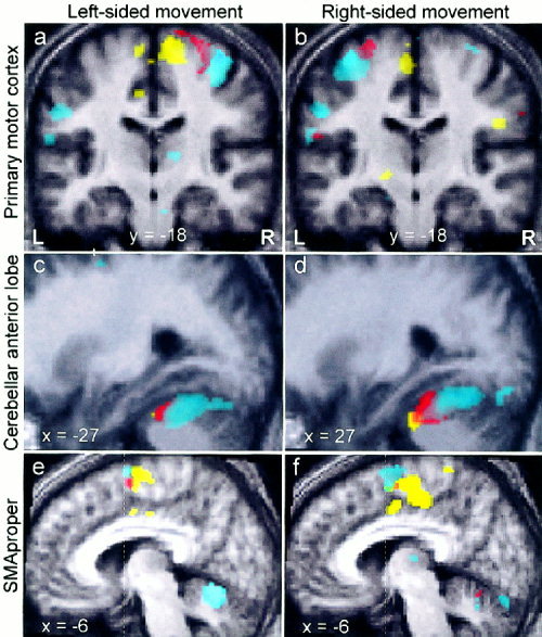
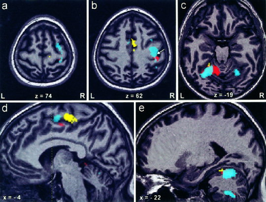
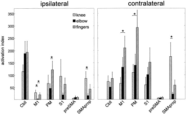
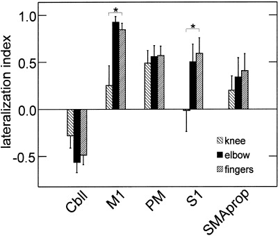
It was shown recently that functional activation across brain motor areas during locomotion and foot movements are similar but differ substantially from activation related to upper extremity movement (Miyai [2001]: Neuroimage 14:1186-1192). The activation pattern may be a function of the behavioral context of the movement rather than of its mechanical properties. We compare motor system activation patterns associated with isolated single-joint movement of corresponding joints in arm and leg carried out in equal frequency and range. Eleven healthy volunteers underwent BOLD-weighted fMRI while performing repetitive elbow or knee extension/flexion. To relate elbow and knee activation to the well-described patterns of finger movement, serial finger-to-thumb opposition was assessed in addition. After identifying task-related voxels using statistical parametric mapping, activation was measured in five regions of interest (ROI; primary motor [M1] and somatosensory cortex [S1], premotor cortex, supplementary motor area [SMA] divided into preSMA and SMA-proper, and cerebellum). Differences in the degree of activation across ROIs were found between elbow and knee movement. SMA-proper activation was prominent for knee, but almost absent for elbow movement (P < 0.05); finger movement produced small but constant SMA-proper activation. Ipsilateral M1 activation was detected during knee and finger movement, but was absent for the elbow task (P < 0.05). Knee movement showed less lateralization in M1 and S1 than other tasks (P < 0.05). The data demonstrate that central motor structures contribute differently to isolated elbow and knee movement. Activation during knee movement shows similarities to gait-related activation patterns.
Copyright 2002 Wiley-Liss, Inc.
Figures
References
-
- Ashe J (1997): Force and the motor cortex. Behav Brain Res 86: 1–15. - PubMed
-
- Babiloni C, Carducci F, Pizzella V, Indovina I, Romani GL, Rossini PM, Babiloni F (1999): Bilateral neuromagnetic activation of human primary sensorimotor cortex in preparation and execution of unilateral voluntary finger movements. Brain Res 827: 234–236. - PubMed
-
- Beisteiner R, Windischberger C, Lanzenberger R, Edward V, Cunnington R, Erdler M, Gartus A, Streibl B, Moser E, Deecke L (2001): Finger somatotopy in human motor cortex. Neuroimage 13: 1016–1026. - PubMed
-
- Boussaoud D (2001): Attention versus intention in the primate premotor cortex. Neuroimage 14: S40–S45. - PubMed
-
- Bushara KO, Wheat JM, Khan A, Mock BJ, Turski PA, Sorenson J, Brooks BR (2001): Multiple tactile maps in the human cerebellum. Neuroreport 12: 2483–2486. - PubMed
Publication types
MeSH terms
LinkOut - more resources
Full Text Sources
