Comparative Study
. 2002 Oct 1;21(19):5007-16.
doi: 10.1093/emboj/cdf526.

The cross-reactive calcium-binding pollen allergen, Phl p 7, reveals a novel dimer assembly

Affiliations
Comparative Study

The cross-reactive calcium-binding pollen allergen, Phl p 7, reveals a novel dimer assembly

Petra Verdino et al. EMBO J. .

Abstract

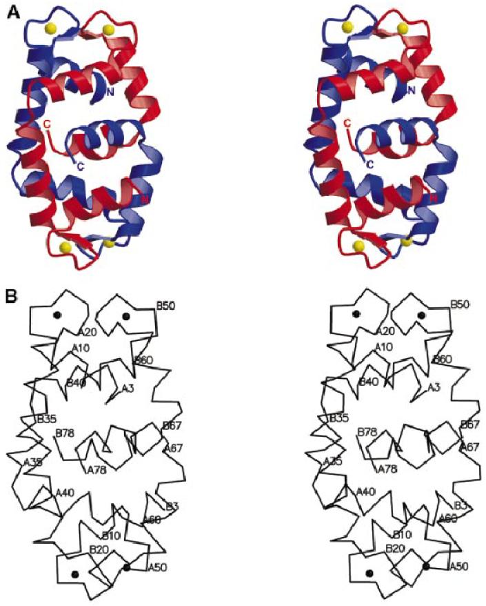
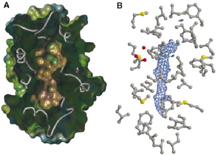
The timothy grass pollen allergen Phl p 7 assembles most of the IgE epitopes of a novel family of 2 EF-hand calcium-binding proteins and therefore represents a diagnostic marker allergen and vaccine candidate for immunotherapy. Here we report the first three-dimensional structure of a representative of the 2 EF-hand allergen family, Phl p 7, in the calcium-bound form. The protein occurs as a novel dimer assembly with unique features: in contrast to well known EF-hand proteins such as calmodulin, parvalbumin or the S100 proteins, Phl p 7 adopts an extended conformation. Two protein monomers assemble in a head-to-tail arrangement with domain-swapped EF-hand pairing. The intertwined dimer adopts a barrel-like structure with an extended hydrophobic cavity providing a ligand-binding site. Calcium binding acts as a conformational switch between an open and a closed dimeric form of Phl p 7. These findings are interesting in the context of lipid- and calcium-dependent pollen tube growth. Furthermore, the structure of Phl p 7 allows for the rational development of vaccine strategies for treatment of sensitized allergic patients.


Figures

None
Fig. 1. (A) Stereo ribbon representation of the intertwined Phl p 7 dimer. Chain A is coloured in blue, chain B is shown in red and the calcium ions are highlighted in yellow. The N-terminal EF-hand of chain A (AI) and the C-terminal EF-hand of chain B (BII) comprise the upper EF-hand pair, whereas the C-terminal EF-hand of chain A (AII) and the N-terminal EF-hand of chain B (BI) form the lower one. The C-terminal Z-helices comprise an equatorial belt. Figures were prepared with MOLSCRIPT (Kraulis, 1991) and rendered using Raster3D (Merritt and Murphy, 1994). (B) Stereo Cα trace of the Phl p 7 dimer, labelled at every tenth amino acid. (C) Sequence alignment of five representative 2 EF-hand pollen allergens (from the top: timothy grass, bermuda grass, olive, alder, birch), the 4 EF-hand pollen allergen Jun o 2 (cypress) and various representative EF-hand proteins. The secondary structure plot is derived from the three-dimensional structure of Phl p 7. The helices of the N- and C-terminal EF-hands (EFI and EFII) and the Z-helix are represented by grey tubes and the short β-strands by black arrows. Gaps are symbolized as dots; hyphens indicate that only parts of the whole sequences are shown. More than 75% homology (conserved exchanges: RK, DE, NQ, TS) within all proteins is shown in red, and residues which exhibit 100% homology within the 2 EF-hand pollen allergens are pictured in blue.
None
Fig. 2. (A) Superimposition of a Phl p 7 monomer with two representative calcium-binding EF-hand proteins. The backbone of Phl p 7 chain A (except the Z-helix) is shown in blue; its calcium ions are pictured in yellow. The calcium-binding loop of the C-terminal EF-hand AII was superimposed with the corresponding loops of the C-terminal EF-hand of a calmodulin domain (1OSA; light green) and S100B (1MHO; orange), respectively. Whereas the two EF-hands of Phl p 7 adopt an extended conformation, the EF-hands of representative EF-hand proteins are arranged in close spatial proximity. In contrast to the rather short three amino acid hinge loop between the calcium-binding sites of Phl p 7, a much longer loop (6–14 residues) allows for the sequential EF-hand pairing in well-known EF-hand proteins. (B) Superimposition of the pairing calcium-binding loops of Phl p 7 and representative EF-hand proteins. The chains of Phl p 7 are shown in blue (AII) and red (BI), respectively, and the calcium ions are represented in yellow. The sequentially pairing EF-hand loops of calmodulin (1OSA), troponin C (1TOP), parvalbumin (1PVB) and S100B (1MHO) are represented in grey. Although in Phl p 7 the pairing occurs between the two monomer chains, the spatial arrangement of the loops is identical to the intrachain paired loops of standard EF-hand proteins.
None
Fig. 3. (A) Stereo diagram of the (3Fo – 2Fc) electron density map at 1.75 Å resolution contoured at 1σ around the 12 amino acid calcium-binding loop of AI (residues 13–24) with the calcium ion (yellow sphere) positioned in the middle. (B) The 7-fold coordination of the calcium ion by oxygen atoms (red) of asparagines or aspartic acids, a peptide carbonyl oxygen, a water molecule and a bidentate glutamic acid in the positions X Y Z –Y –X –Z according to the nomenclature of Kretsinger (Kretsinger and Nockolds, 1973; Lewit-Bentley and Rety, 2000). (C) The loop pairing interactions between AI (blue) and BII (red): two hydrogen bonds (green dotted lines) are formed between the peptide planes of I20 (chain A) and I55 (chain B). Furthermore, this short β-sheet assembly is stabilized by hydrophobic interactions (red dotted lines) between the side chains of I20 and I55 as well as K19 and F54. The latter residues are also involved in the calcium coordination via their backbone carbonyl oxygen atoms [in position –Y in (B)].
None
Fig. 4. (A) pH-dependent size exclusion chromatography. The protein standard is shown as a black dotted line. Phl p 7 assembles as a dimer with an apparent mol. wt of 14 kDa at both pH 7.1 (green) and pH 4.7 (blue). (B) Size exclusion chromatography coupled to ICP-MS (inductively coupled plasma mass spectrometry). The UV absorption traces at 210 nm (coloured lines) and the corresponding online calcium ion count mass spectrometry traces (black lines) are shown. The native Phl p 7 protein and the heat-treated sample after re-annealing in the presence of calcium yield identical elution profiles; the coincident traces are coloured in blue. The heat-treated protein refolded in the presence of EGTA is pictured in red. Room temperature incubation of Phl p 7 in the presence of EGTA is shown in green. The yellow trace represents the protein after heat denaturation and refolding in the presence of EGTA followed by subsequent addition of excessive CaCl2. Phl p 7 remains dimeric under all applied conditions, even in the completely calcium-free form. Calcium depletion results in a shorter elution time, corresponding to an increase of the hydrodynamic radius of the dimer. Addition of calcium to the EGTA-treated protein leads to re-incorporation of the metal ion and an identical elution time as for the untreated calcium-bound protein.
None
Fig. 5. (A) Intersection through the Phl p 7 dimer. The polypeptide backbone is represented in light grey. Surfaces were calculated and coloured according to their hydrophobicity, from brown (hydrophobic) through green to blue (hydrophilic) with SYBYL® 6.7.1 (Tripos Inc.). The protein’s outer surface is hydrophilic (especially in the calcium-binding regions), whereas the cavity is predominantly hydrophobic and completely sealed by hydrophobic side chains. (B) Zooming into the hydrophobic cavity: the (3Fo – 2Fc) electron density contoured at 1σ of the ligand (blue map) surrounded by the lining residues. Eight isoleucine, eight leucine, two alanine and one valine residue comprise the lateral walls of the cavity, whereas clusters of phenylalanine residues form the top and the bottom. The only polar side chain within the hydrophobic assembly, an aspartic acid coordinating a solvent molecule, is located in the more globular upper part of the cavity. (C and D) Surface plots of key features of the Phl p 7 dimer assembly: the ridge evolving at the ‘kink’ region and the calcium-coordinating site AI/BII. The left pictures represent the polypeptide backbones of chain A (blue) and chain B (red). The electrostatic potential distribution coloured from red (negative) through white to blue (positive) with GRASP (Nicholls et al., 1991) is shown in the middle. The right pictures show the surface hydrophobicity that is colour-coded analogously to (A).

Similar articles

Cited by

References

    1. Babu Y.S., Bugg,C.E. and Cook,W.J. (1988) Structure of calmodulin refined at 2.2 Å resolution. J. Mol. Biol., 204, 191–204. - PubMed
    1. Batanero E., Villalba,M., Ledesma,A., Puente,X.S. and Rodriguez,R. (1996) Ole e 3, an olive-tree allergen, belongs to a widespread family of pollen proteins. Eur. J. Biochem., 241, 772–778. - PubMed
    1. Bennett M.J., Schlunegger,M.P. and Eisenberg,D. (1995) 3D domain swapping: a mechanism for oligomer assembly. Protein Sci., 4, 2455–2468. - PMC - PubMed
    1. Brewbaker J.L. and Kwack,B.H. (1963) The essential role of calcium ion in pollen germination and pollen tube growth. Am. J. Bot., 50, 859–865.
    1. Brünger A.T. et al. (1998) Crystallography and NMR system: a new software suite for macromolecular structure determination. Acta Crystallogr. D, 54, 905–921. - PubMed

Publication types

Associated data