. 2002 Oct 1;21(19):5017-25.
doi: 10.1093/emboj/cdf529.

The human scavenger receptor class B type I is a novel candidate receptor for the hepatitis C virus

Affiliations

The human scavenger receptor class B type I is a novel candidate receptor for the hepatitis C virus

Elisa Scarselli et al. EMBO J. .

Abstract

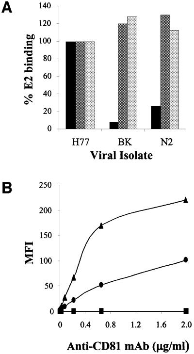
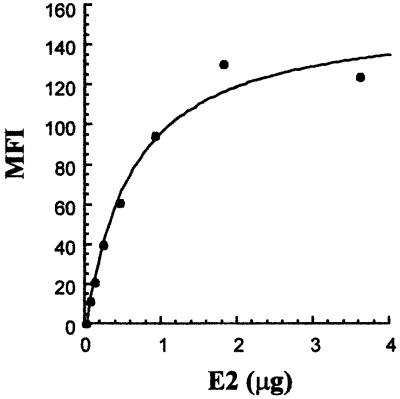
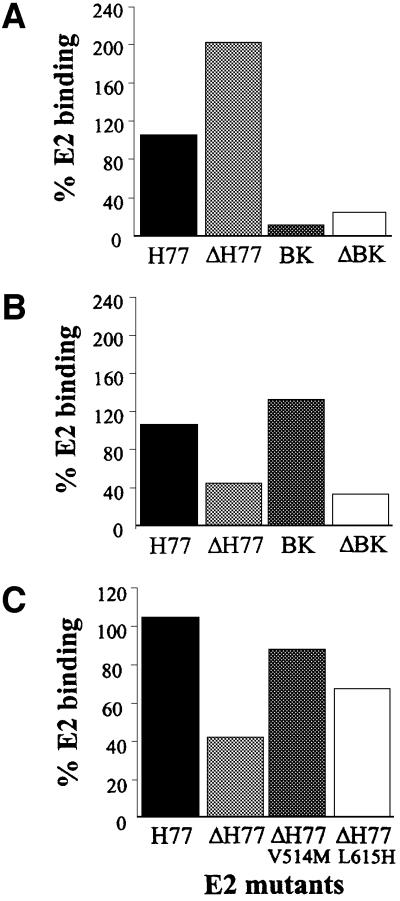
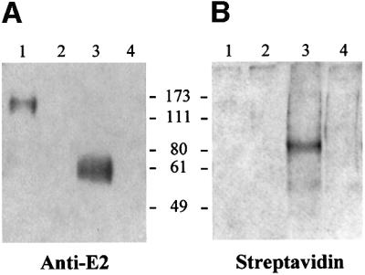
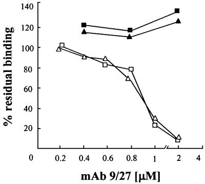
We discovered that the hepatitis C virus (HCV) envelope glycoprotein E2 binds to human hepatoma cell lines independently of the previously proposed HCV receptor CD81. Comparative binding studies using recombinant E2 from the most prevalent 1a and 1b genotypes revealed that E2 recognition by hepatoma cells is independent from the viral isolate, while E2-CD81 interaction is isolate specific. Binding of soluble E2 to human hepatoma cells was impaired by deletion of the hypervariable region 1 (HVR1), but the wild-type phenotype was recovered by introducing a compensatory mutation reported previously to rescue infectivity of an HVR1-deleted HCV infectious clone. We have identified the receptor responsible for E2 binding to human hepatic cells as the human scavenger receptor class B type I (SR-BI). E2-SR-BI interaction is very selective since neither mouse SR-BI nor the closely related human scavenger receptor CD36, were able to bind E2. Finally, E2 recognition by SR-BI was competed out in an isolate-specific manner both on the hepatoma cell line and on the human SR-BI-transfected cell line by an anti-HVR1 monoclonal antibody.


Figures

None
Fig. 1. (A) Histograms showing the E2 binding of different viral isolates to human cell lines as measured by FACS analysis. Values are expressed as a percentage of the H77-E2 isolate binding. Molt-4 cells are represented by the black histogram. The human hepatic cell line Huh7 is represented by the dark gray histogram and the HepG2 cells by the light gray histogram. (B) Cell surface expression of CD81 measured on the different cell lines by FACS analysis with the anti-CD81 (mAb1.3.3.22) antibody in a direct binding assay. Molt-4 cells are represented by the triangle, Huh7 by the circle and HepG2 by the square. On the y axis net median fluorescence intensity (MFI) values are reported.
None
Fig. 2. Binding saturation curve of H77-E2 recombinant protein to HepG2 cells. On the y axis, net median fluorescence intensity (MFI) values were calculated by subtracting the background MFI from non-specific binding of primary and secondary antibodies to the values obtained with E2. On the x axis, the monomeric E2 content was calculated as described in Material and methods.
None
Fig. 3. (A) A histogram showing the binding of the E2 recombinant proteins deleted of the HVR1 to Molt-4. (B) A histogram showing the binding of the E2 recombinant proteins deleted of the HVR1 to HepG2. (C) A histogram showing the binding of the E2 recombinant proteins deleted of the HVR1 with compensatory mutations to HepG2. E2 binding is measured by FACS analysis and values are expressed as a percentage of the H77-E2 isolate binding.
None
Fig. 4. (A) Western blot detection of cell surface-bound E2. Biotinylated HepG2 cells were incubated in the presence (lanes 1 and 3) or in the absence (lanes 2 and 4) of E2 recombinant protein. Bound E2 was cross-linked with DTSSP and the E2–receptor complexes were immunoprecipitated with an antibody against the His tag of the E2 recombinant protein. Samples eluted under both non-reducing (lanes 1 and 2) and reducing (lanes 3 and 4) conditions were loaded onto a 10% SDS–PAGE gel. E2 protein was detected as a monomer under reducing conditions (lane 3) and at a higher molecular weight under non- reducing conditions (lane 1), by using an anti-E2 rat mAb followed by anti-rat HRP-conjugated. (B) Immunoblot detection of biotinylated cell surface proteins interacting with E2. The reactivity with HRP conjugated streptavidin revealed a biotinylated protein band at 82 kDa only under reducing conditions (lane 3).
None
Fig. 5. (A) Silver staining of an SDS–PAGE gel loaded with samples obtained after the purification step with Con-A–Sepharose and deglycosylation with the PNGase F. The arrows show the purified receptor migrating at 82 kDa (lane 1) before PNGase F treatment (–), and migrating at 54 kDa (lane 2) after deglycosylation (+). In control samples cross-linking was performed in the absence of E2 (lanes 3 and 4). (B) Western blot detection of the glycosylated (lane 1) and deglycosylated (lane 2) receptor protein by incubation with rabbit anti-SR-BI polyclonal antibodies followed by HRP-conjugated anti-rabbit. Lanes 3 and 4 represent the control experiments performed in the absence of E2.
None
Fig. 6. (A) FACS analysis of anti-SR-BI binding to CHO transfected cells. Transfection was performed with pcDNA3, pcDNA3-hSR-BI or pcDNA3-mSR-BI. (B) FACS analysis of E2-H77 binding to CHO transfected cells.
None
Fig. 7. FACS analysis showing the binding of E2 recombinant proteins derived from H77 and BK isolates to CHO wild type (unshaded continuous curve), CHO-CD36 (unshaded dashed curve) and CHO-SR-BI (shaded curve).
None
Fig. 8. A histogram showing the binding of the E2 recombinant proteins deleted of the HVR1 to CHO-SR-BI transfected cells. Binding is measured by FACS analysis and values are expressed as a percentage of the H77-E2 isolate binding.
None
Fig. 9. Competition of E2 binding to HepG2 cells and CHO-SR-BI by the anti-HVR1 mAb 9/27 reactive with H77-E2. Binding was detected by FACS analysis and is expressed as a percentage of the MFI values obtained in the absence of competitor. H77-E2 binding to HepG2 (open triangle) and CHO-SR-BI (open square). BK-E2 binding to HepG2 (filled triangle) and to CHO–SR-BI (filled square).

Similar articles

Cited by

References

    1. Acton S.L., Scherer,P.E., Lodish,H.F. and Krieger,M. (1994) Expression cloning of SR-BI, a CD36-related class B scavenger receptor. J. Biol. Chem., 269, 21003–21009. - PubMed
    1. Acton S., Rigotti,A., Landschulz,K.T., Xu,S., Hobbs,H.H. and Krieger,M. (1996) Identification of scavenger receptor SR-BI as a high density lipoprotein receptor. Science, 271, 518–520. - PubMed
    1. Agnello V., Abel,G., Elfahal,M., Knight,G.B. and Zhang,Q.X. (1999) Hepatitis C virus and other flaviviridae viruses enter cells via low density lipoprotein receptor. Proc. Natl Acad. Sci. USA, 96, 12766–12771. - PMC - PubMed
    1. Allander T., Forns,X., Emerson,S.U., Purcell,R.H. and Bukh,J. (2000) Hepatitis C virus envelope protein E2 binds to CD81 of tamarins. Virology, 277, 358–367. - PubMed
    1. Babitt J., Trigatti,B., Rigotti,A., Smart,E.J., Anderson,R.G., Xu,S. and Krieger,M. (1997) Murine SR-BI, a high density lipoprotein receptor that mediates selective lipid uptake, is N-glycosylated and fatty acylated and colocalizes with plasma membrane caveolae. J. Biol. Chem., 272, 13242–13249. - PubMed

Publication types

MeSH terms