. 2002 Oct 1;21(19):5097-108.
doi: 10.1093/emboj/cdf512.

Two distinct phosphoinositide 3-kinases mediate polypeptide growth factor-stimulated PKB activation

Affiliations

Two distinct phosphoinositide 3-kinases mediate polypeptide growth factor-stimulated PKB activation

Alexandre Arcaro et al. EMBO J. .

Abstract

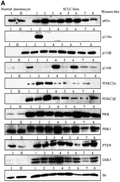
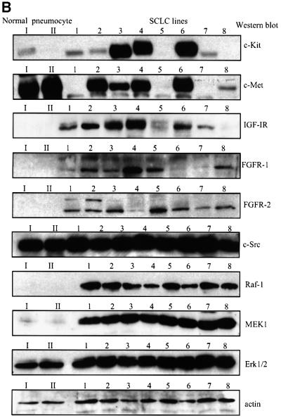
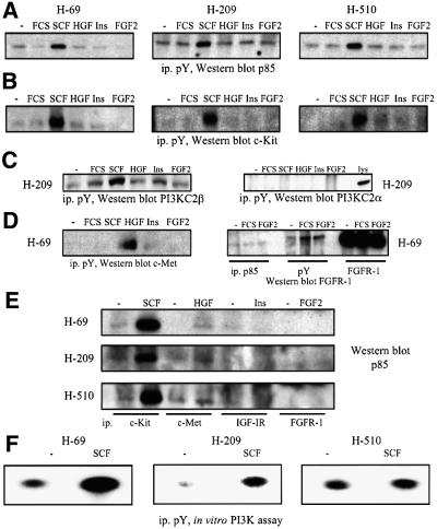
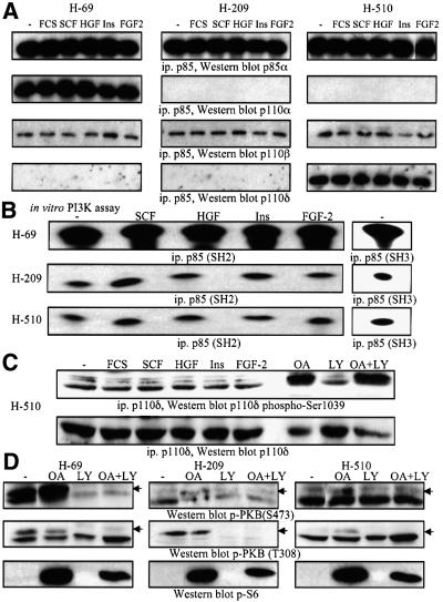
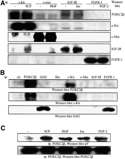
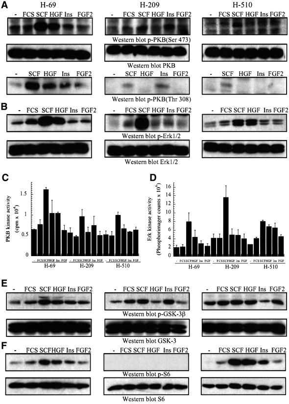
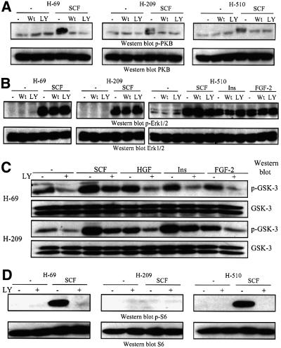
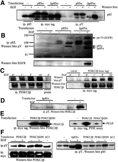
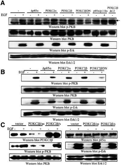
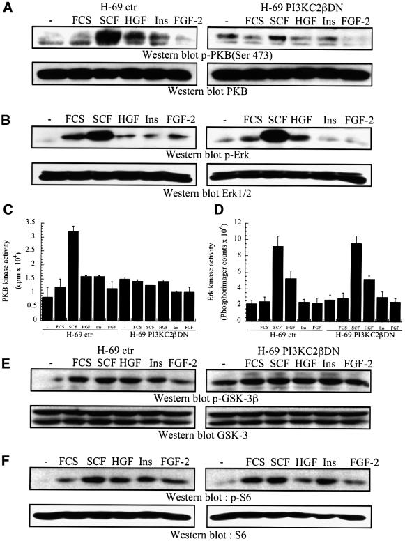
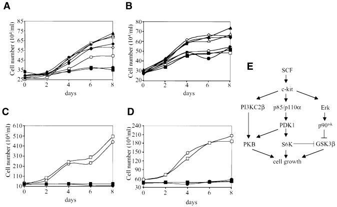
Eight human isoforms of phosphoinositide 3-kinases (PI3Ks) exist, but their individual functions remain poorly understood. Here, we show that different human small cell lung carcinoma (SCLC) cell lines overexpress distinct subsets of class I(A) and II PI3Ks, which results in striking differences in the signalling cascades activated by stem cell factor (SCF). Over expression of class I(A) p85/p110alpha in SCLC cells increased SCF-stimulated protein kinase B (PKB) activation and cell growth, but did not affect extracellular signal-regulated kinase (Erk) or glycogen synthase kinase-3 (GSK-3). This effect was selective, since it was not observed in SCLC cell lines overexpressing p85/p110beta or p85/p110delta. The SCF receptor associated with both class I(A) p85 and class II PI3KC2beta, and both enzymes contributed to SCF-stimulated PKB activity. A dominant-negative PI3KC2beta blocked both PKB activation and SCLC cell growth in response to SCF. Together our data provide novel insights into the specificity and functional significance of PI3K signalling in human cancer.


Figures

None
Fig. 1. Human SCLC cell lines overexpress different isoforms of class IA and II PI3Ks. (A) and (B) Purified human type II pneumocytes from normal lung tissue (I, II) or human SCLC cell lines (1–8) were lysed and detergent-soluble fractions analysed by western blotting with antibodies specific for various PI3K isoforms (A), polypeptide growth factor receptors (B) or the proteins indicated (A and B). The SCLC cell lines analysed were: 1, HC-33; 2, H-69; 3, H-209; 4, H-510; 5, H-524; 6, H-1045; 7, H-1622; 8, H-2171.
None
Fig. 2. Polypeptide growth factor stimulation of SCLC cell lines induces the recruitment of p85 to phosphotyrosine-containing signalling complexes. (AD) SCLC cells were treated with SCF (10 ng/ml), HGF (10 ng/ml), insulin (3 µg/ml) or FGF-2 (1 ng/ml) for 5 min at 37°C. Anti-phosphotyrosine immunoprecipitates were analysed by western blotting with antibodies specific for the proteins indicated. In some cases lysates of the respective SCLC cell lines were analysed in parallel as positive controls (lys). (E) Lysates from SCLC cells prepared as in (A) were immunoprecipitated with antibodies against different receptors, and samples analysed by western blotting with anti-p85 antibodies. (F) SCLC cells (as indicated) were treated with SCF (10 ng/ml) for 5 min at 37°C. Anti-phosphotyrosine immunoprecipitates were assayed for in vitro PI3K activity. Radioactive phospholipids were separated by thin layer chromatography, and spots visualized by autoradiography.
None
Fig. 3. SCLC cells overexpressing p110α display higher p85-associated PI3K activity than cells overexpressing p110β or p110δ. (A) and (B) SCLC cells (as indicated) were treated with SCF (10 ng/ml), HGF (10 ng/ml), insulin (3 µg/ml) or FGF-2 (1 ng/ml) for 5 min at 37°C. Anti-p85 immunoprecipitates (A) were analysed by western blotting with antibodies specific for class IA PI3Ks. (B) Anti-p85 immunoprecipitates prepared with antibodies to the SH2 or SH3 domain were assayed for in vitro PI3K activity as in Figure 2. (C) Lysates from H-510 cells stimulated as in (A) and (B) were analysed by western blotting with phospho-specific antibodies to Ser1039 of p110δ or anti-p110δ antiserum. Lysates from H-510 cells pre-treated for 2 h with vehicle (–), LY294002 (LY, 10 µM), okadaic acid (OA, 1 µM) or LY294002 and OA, were analysed in parallel. (D) Lysates from SCLC cells as in (C) were analysed by western blotting with antibodies specific for activated PKB (p-PKB, Ser473 or T308), or Ser240/244-phosphorylated S6 protein (p-S6).
None
Fig. 4. Polypeptide growth factor regulation of the class II PI3KC2β in human SCLC cells. (A) H-209 cells were treated with SCF (10 ng/ml), HGF (10 ng/ml), insulin (3 µg/ml) or FGF-2 (1 ng/ml) for 5 min at 37°C. The samples were immunoprecipitated with antibodies specific for various receptors and analysed by western blotting with antibodies specific for PI3KC2β or the receptors, as indicated. (B) Immuno precipitates from resting H-69 cells prepared with control antiserum (ctr), or antibodies specific for the proteins indicated were analysed by western blotting with antibodies specific for PI3KC2β or the receptors and adapters indicated. (C) H-209 cells were treated as in (A), and anti-PI3KC2β immunoprecipitates analysed by western blotting with antibodies specific for phosphotyrosine (pY) or PI3KC2β.
None
Fig. 5. Differential overexpression of PI3K isoforms affects downstream signalling cascades in human SCLC cells. (A–F) SCLC cells were treated with SCF (10 ng/ml), HGF (10 ng/ml), insulin (3 µg/ml), or FGF-2 (1 ng/ml) for 5 min or (F) 10 min, at 37°C. (A and B) and (E and F) The samples analysed by western blotting with antibodies specific for (A) activated PKB (p-PKB, Ser473 or Thr308) or total PKB, (B) activated Erk1/2 (p-Erk1/2) or total Erk, (E) Ser9-phosphorylated GSK-3β (p-GSK-3β) or total GSK-3, (F) Ser240/244-phosphorylated S6 protein (p-S6) or total S6 protein. Alternatively, anti-PKB (C) or anti-Erk (D) immunoprecipitates were assayed for in vitro protein kinase activity, using crosstide (C) or myelin basic protein (D) as substrates.
None
Fig. 6. Pharmacological PI3K inhibitors abrogate polypeptide growth factor-stimulated PKB activation in human SCLC cells. (A–D) SCLC cells were pre-treated with vehicle (–), wortmannin (Wt, 50 nM) or LY294002 (LY, 10 µM) for 15 min at 37°C. (AC) The cells were then stimulated with SCF (10 ng/ml), HGF (10 ng/ml), insulin (3 µg/ml) or FGF-2 (1 ng/ml) for 5 min or (D) 10 min, at 37°C. After stimulation the cells were lysed, and samples analysed by western blotting with antibodies specific for (A) activated PKB (p-PKB) or total PKB, (B) activated Erk1/2 (p-Erk1/2) or total Erk, (C) Ser9- phosphorylated GSK-3β (p-GSK-3β) or total GSK-3, (D) Ser240/244-phosphorylated S6 protein (p-S6) or total S6 protein.
None
Fig. 7. Evaluation of the selectivity of dominant-negative PI3K constructs. (A) COS-7 cells transiently transfected with the constructs encoding myc-tagged p85α or Δp85α were stimulated with EGF, and anti-p85α or anti-phosphotyrosine immunoprecipitates analysed by western blotting with anti-p85 antibodies. (B) The blot was reprobed with anti-phosphotyrosine (top) or anti-EGFR (bottom) antibodies. The position of myc-tagged p85α and Δp85α, and the pp170 band corresponding to the EGFR are shown. (C) 293 cells (left) or 293 cells transfected with a construct encoding myc-tagged PI3KC2β (right) were stimulated with EGF for various lengths of time. Samples were immunoprecipitated with anti-PI3KC2β antiserum, pre-immune serum (left) or anti-myc tag antibodies (right), and analysed by western blotting with anti-phosphotyrosine (pY) or anti-PI3KC2β antibodies. (D) COS-7 cells were transiently transfected with the Δp85α construct and stimulated with EGF. Anti-phosphotyrosine immunoprecipitates were analysed by western blotting with anti-PI3KC2β antiserum. (E) 293 cells were transfected with a construct encoding myc-tagged PI3KC2β wild-type or DN. Anti-myc tag immunoprecipitates were analysed by western blotting with anti-PI3KC2β antiserum (left) or assayed for in vitro PI3K activity (right). (F) 293 cells transfected with the cDNA constructs indicated were stimulated with EGF. Anti- phosphotyrosine or anti-myc tag immunoprecipitates were analysed by western blotting with anti-PI3KC2β antiserum or with anti-p85 antibodies.
None
Fig. 8. Transfection of class IA or II PI3Ks differentially affects downstream signalling cascades in mammalian cells. (A) HEK293 or (B) COS cells were transiently transfected with various PI3K constructs (as indicated). Serum-starved cells were stimulated with EGF (50 nM) for 5 min at 37°C, and samples analysed by western blotting for activated PKB (p-PKB), activated Erk1/2 (p-Erk1/2) or total respective proteins. (C) 293 cells were transfected with antisense PI3KC2β construct (as), wild-type PI3KC2β (s) or empty vector. Serum-starved cells were stimulated with EGF (50 nM) for 5 min at 37°C, and samples analysed by western blotting for PI3KC2β, p85α, activated PKB (p-PKB), activated Erk1/2 (p-Erk1/2) or total respective proteins.
None
Fig. 9. Transfection of kinase-inactive PI3KC2β selectively inhibits PKB activity. (A–F) H-69 cells transfected with an empty vector or PI3KC2βDN were treated with SCF (10 ng/ml), HGF (10 ng/ml), insulin (3 µg/ml) or FGF-2 (1 ng/ml) for 5 min, or (F) 10 min, at 37°C. (A and B) and (E and F) Samples were analysed by western blotting with antibodies specific for (A) activated PKB (p-PKB Thr308) or total PKB, (B) activated Erk1/2 (p-Erk1/2) or total Erk, (E) Ser9-phosphorylated GSK-3β (p-GSK-3β) or total GSK-3, (F) Ser240/244-phosphorylated S6 protein (p-S6) or total S6 protein. Alternatively, anti-PKB (C) or anti-Erk (D) immunoprecipitates were assayed for in vitro protein kinase activity, using crosstide (C) or myelin basic protein (D) as substrates.
None
Fig. 10. SCF-stimulated SCLC cell growth is dependent on PI3K signalling. (A) H-69 cells or (B) H-69 transfected with PI3KC2βDN were treated with SCF (10 ng/ml, squares), insulin (3 µg/ml, triangles), FGF-2 (1 ng/ml, diamonds) or no growth factor (circles) in the presence (closed symbols) or absence (open symbols) of LY294002 (10 µM) and cell number determined by counting every 2 days. (C) H-209 or (D) H-510 cells were treated with SCF (squares) or no growth factor (circles) in the presence (closed symbols) or absence (open symbols) of LY294002 and cell number determined by counting every 2 days. The growth factors and inhibitor were added at 0 and 4 days. (E) Schematic representation of the PI3K-dependent signalling pathways involved in SCLC cell growth in response to SCF.

Similar articles

Cited by

References

    1. Alessi D.R., James,S.R., Downes,C.P., Holmes,A.B., Gaffney,P., Reece,C. and Cohen,P. (1997) Purification and characterization of a phosphatidylinositol 3,4,5 trisphosphate-dependent protein kinase (PDK1) which phosphorylates and activates protein kinase Bα. Curr. Biol., 7, 261–269. - PubMed
    1. Alessi D.R., Kozlowski,M.T., Weng,Q.P., Morrice,N. and Avruch,J. (1998) 3-Phosphoinositide-dependent protein kinase 1 (PDK1) phosphorylates and activates the p70 S6 kinase in vivo and in vitro. Curr. Biol., 8, 69–81. - PubMed
    1. Arcaro A., Volinia,S., Zvelebil,M.J., Stein,R., Watton,S.J., Layton,M.J., Gout, I., Ahmadi, K., Downward,J. and Waterfield,M.D. (1998) Human phosphoinositide 3-kinase C2β, the role of calcium and the C2 domain in enzyme activity. J. Biol. Chem., 273, 33082–33090. - PubMed
    1. Arcaro A., Zvelebil,M.J., Wallasch,C., Ullrich,A., Waterfield,M.D. and Domin,J. (2000) Class II phosphoinositide 3-kinases are downstream targets of activated polypeptide growth factor receptors. Mol. Cell. Biol., 20, 3817–3830. - PMC - PubMed
    1. Beeton C.A., Chance,E.M., Foukas,L.C. and Shepherd,P.R. (2000) Comparison of the kinetic properties of the lipid- and protein-kinase activities of the p110α and p110β catalytic subunits of class-Ia phosphoinositide 3-kinases. Biochem. J., 350, 353–359. - PMC - PubMed

Publication types

MeSH terms