. 2002 Oct 1;21(19):5109-17.
doi: 10.1093/emboj/cdf509.

A novel vasopressin-induced transcript promotes MAP kinase activation and ENaC downregulation

Affiliations

A novel vasopressin-induced transcript promotes MAP kinase activation and ENaC downregulation

Marie Nicod et al. EMBO J. .

Abstract

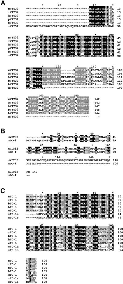
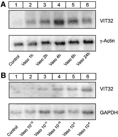
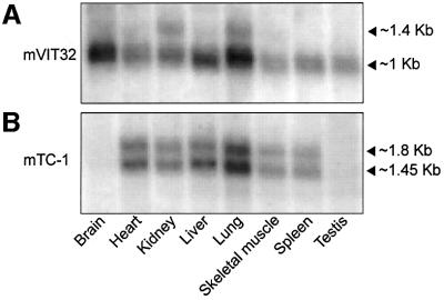
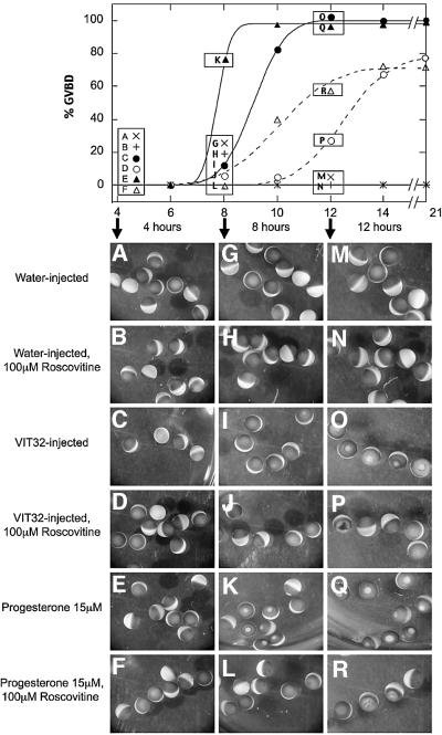
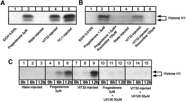
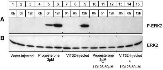
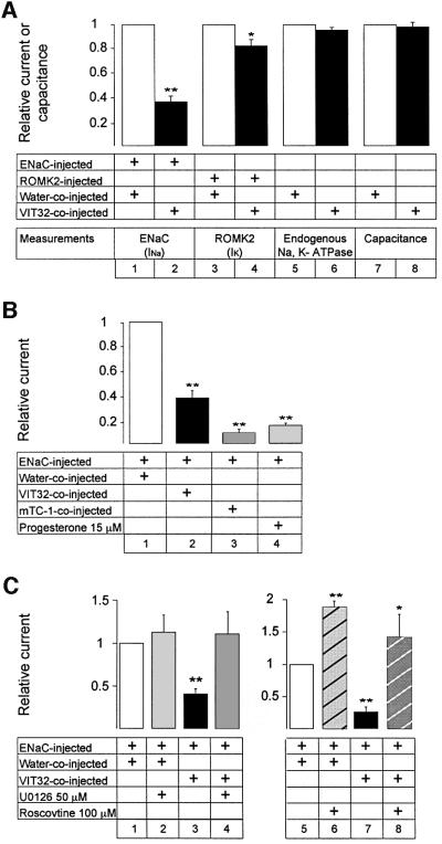
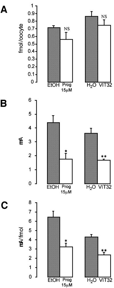
In the principal cell of the renal collecting duct, vasopressin regulates the expression of a gene network responsible for sodium and water reabsorption through the regulation of the water channel and the epithelial sodium channel (ENaC). We have recently identified a novel vasopressin-induced transcript (VIT32) that encodes for a 142 amino acid vasopressin-induced protein (VIP32), which has no homology with any protein of known function. The Xenopus oocyte expression system revealed two functions: (i) when injected alone, VIT32 cRNA rapidly induces oocyte meiotic maturation through the activation of the maturation promoting factor, the amphibian homolog of the universal M phase trigger Cdc2/cyclin; and (ii) when co-injected with the ENaC, VIT32 cRNA selectively downregulates channel activity, but not channel cell surface expression. In the kidney principal cell, VIP32 may be involved in the downregulation of transepithelial sodium transport observed within a few hours after vasopressin treatment. VIP32 belongs to a novel gene family ubiquitously expressed in oocyte and somatic cells that may be involved in G to M transition and cell cycling.


Figures

None
Fig. 1. (A) Alignment of ortholog VIT32 proteins from mouse, rat, human, pig, bovine and frog (Xenopus laevis). Mouse (m), rat (r) and human (h) VIT32 amino acid sequences are available at the DDBJ/EMBL/GenBank databases as hypothetical proteins with unknown function (accession Nos: BAB26163, NP_599200 and NP_068378, respectively). Pig (p), bovine (b) and Xenopus laevis (x) VIT32 sequences were obtained by alignment of multiple ESTs. (B) Comparison of amino acid sequences of mVIT32 and mTC-1. The mTC-1 amino acid sequence is available at the DDBJ/EMBL/GenBank databases under accession No. BAB25041. (C) Alignment of ortholog TC-1 proteins from mouse, rat, bovine, chicken, human and zebra fish. The hTC-1 amino acid sequence is available at the DDBJ/EMBL/GenBank databases under accession No. NP_064515. Rat (r), bovine (b), chicken (c) and zebra fish (z) TC-1 sequences were obtained by alignment of multiple ESTs.
None
Fig. 2. Vasopressin regulates mVIT32 mRNA abundance in mpkCCD cells. (A) Time course. Northern blot analysis with mVIT32 probe was performed on mRNAs extracted from untreated mpkCCD cells (control) or vasopressin (10–8 M)-stimulated mpkCCD cells after indicated period of time. (B) Dose response. Northern blot analysis was performed on mRNAs extracted from untreated mpkCCD cells (control) or mpkCCD cells stimulated for 4 h with vasopressin at different concentrations.
None
Fig. 3. (A) Northern blot analysis of mVIT32 mRNA expression in mouse tissues. In the majority of tested tissues (brain, heart, liver, skeletal muscle, spleen, testis), a single mVIT32 transcript of 1 kb long is present. In kidney and lung, an additional minor transcript of 1.4 kb long is expressed. (B) Northern blot analysis of mTC-1 mRNA expression in mouse tissues. In most of the tested tissues (heart, kidney, liver, lung, skeletal muscle and spleen), two mTC-1 transcripts of 1.45 and 1.8 kb long are present. In brain and testis, no TC-1 expression was detected.
None
Fig. 4. Induction of oocyte maturation by mVIT32. GVBD is scored by the appearance of a white spot on the oocyte animal pole reflecting maturation. Water-injected oocytes incubated with DMSO (0.5%) (x on the top panel; A, G and M) or with roscovitine (100 µM) (+ on the top panel; B, H and N) show no maturation. GVBD was delayed by 2 h for oocytes injected with mVIT32 cRNA (filled circles on the top panel; C, I and O), compared with progesterone (15 µM)-incubated oocytes (filled triangles on the top panel; E, K and Q). In presence of roscovitine (100 µM), mVIT32- (open circle on the top panel; D, J and P) and progesterone-induced (open triangle on the top panel; F, L and R) maturation was delayed by 4 h. At 12 h, mVIT32- and progesterone-induced maturation had apparently different morphologic features, depending on the presence of roscovitine. The percentage of oocytes with GVBD as a function of time after treatment was evaluated in groups of 38–40 oocytes. The best fitting cumulative distribution functions are shown as lines. The parameters of these functions are indicated in the text.
None
Fig. 5. Kinase activity of Cdc2–cyclin B complexes. (A) mVIT32 and mTC-1 cRNAs injection induce Cdc2 activity in Xenopus oocytes. Cdc2 activity in total Xenopus oocytes protein extracts was tested by histone H1 phosphorylation assay (see Materials and methods). Oocyte protein extracts were prepared either from oocytes treated for 13 h with EtOH (0.03%) or progesterone (3 µM) or from oocytes incubated for 13 h after water or mVIT32 cRNA injection. Protein extracts prepared from EtOH-treated (lane 1) and water-injected oocytes (lane 3) show no phosphorylation of histone H1. Progesterone treatment (lane 2) as well as mVIT32 (lane 4) or mTC-1 (lane 5) cRNA injection leads to induction of histone H1 phosphorylation. This experiment was repeated on three independent batches of oocytes with similar results. (B) Roscovitine inhibits the mVIT32-induced Cdc2 activity. Roscovitine (100 µM) was added to oocytes simultaneously with progesterone or immediately after cRNA injection. The kinase assay was performed 13 h after progesterone addition or mVIT32 cRNA injection. Ethanol-treated (lane 1) and water-injected oocytes (lane 4) show no phosphorylation of histone H1. Progesterone-induced kinase activity (lane 2) is fully inhibited by roscovitine (lane 3). mVIT32-induced kinase activity (lane 5) is fully inhibited by roscovitine (lane 6). (C) UO126 inhibits the mVIT32- and progesterone-induced kinase activity. Oocytes were incubated with or without UO126 (50 µM) 12 h before progesterone stimulation or cRNA injection. UO126 (50 µM) was also present in the oocyte incubating medium after progesterone stimulation or cRNA induction. The kinase activity was measured 0, 6 and 12 h after progesterone stimulation or cRNA injection. Water-injected oocytes (lanes 1–3) show no phosphorylation of histone H1. Progesterone (lanes 4–6) induces kinase activity as early as 6 h after treatment. mVIT32- (lanes 7–9) induced histone H1 phosphorylation is delayed and occurs after 12 h (lane 9). UO126 fully inhibits progesterone (lanes 10–12), and mVIT32 (lanes 13–15) induced kinase activity.
None
Fig. 6. mVIT32 injection and progesterone treatment induce ERK2 phosphorylation. Immunoblotting with antibodies directed against ERK1/2 and phospho-ERK1/2 was performed on the same oocyte protein extracts as those used for detection of Cdc2 kinase activity (Figure 5C). Water-injected oocytes (lanes 1–3) show no phosphorylation of ERK2. Progesterone- (lanes 4–6) and mVIT32- (lanes 7–9) induced ERK2 phosphorylation was fully inhibited by UO126 (lanes 10–15). mVIT32-induced ERK2 phosphorylation is delayed compared to the progesterone effect.
None
Fig. 7. For all conditions, 4–8 experiments were performed with at least five oocytes measured per condition. Oocytes were injected with 1 ng of cRNAs of each α, β and γ subunit of ENaC or with 0.2 ng of ROMK2 cRNA. About 12 h later, oocytes were co-injected with water, mVIT32 or mTC-1 cRNA, or treated with progesterone. Five hours later, electrophysiological measurements were made to test ion channel macroscopic currents (INa and IK), endogenous Na,K-ATPase activity and membrane capacitance. (A) Effect of mVIT32 on ENaC and ROMK2 currents, on oocyte capacitance and on endogenous Na,K-ATPase activity. The absolute values for ENaC, ROMK2 and the endogenous Na,K-ATPase currents was 917 ± 297 nA, 475 ± 43 nA and 65 ± 10 nA, respectively. The absolute value of oocyte capacitance was 274 ± 21 nF. mVIT32 significantly decreases INa (lane 1 versus 2) with little effect on IK (lane 3 versus 4). mVIT32 has no effect on endogenous Na,K-ATPase activity (lane 5 versus 6) and oocyte membrane capacitance (lane 7 versus 8). (B) Effect of mVIT32, mTC-1 and progesterone on ENaC current. mVIT32 (lane 2), mTC-1 (lane 3) and progesterone (lane 4) downregulate ENaC activity (lane 1). (C) U0126 and roscovitine inhibit the effect of mVIT32 on ENaC current. UO126 (lane 4) and roscovitine (lane 8) restore the sodium current downregulated by mVIT32 (lanes 3 and 7). *p < 0.05; **p < 0.001 (Student’s t-test).
None
Fig. 8. Effect of mVIT32 and progesterone on ENaC current and ENaC cell surface expression. Oocytes were injected with 2 ng of each α, β and γ subunit of ENaC cRNA. Twelve hours later, these oocytes were injected with 3 ng of mVIT32 cRNA or with the same volume of water, or treated with either 15 µM progesterone or ethanol (0.15%). Eight hours later, cell surface expression of ENaC was measured. INa was measured on the same oocytes 1 h after measurement of ENaC cell surface expression. In each group, ENaC cell surface expression and INa were measured on 30–36 oocytes. (A) Cell surface expression of ENaC. Neither progesterone nor mVIT32 affect cell surface expression of ENaC. (B) ENaC current. INa was significantly decreased under progesterone and mVIT32 effects. (CINa:ENaC cell surface expression ratio. This ratio is proportional to the total channel open probability and is significantly decreased by progesterone and mVIT32. NS, not significant; *p < 0.05; **p < 0.001 (Student’s t-test).

Similar articles

Cited by

References

    1. Bankir L. (2001) Antidiuretic action of vasopressin: quantitative aspects and interaction between V1a and V2 receptor-mediated effects. Cardiovasc. Res., 51, 372–390. - PubMed
    1. Bayaa M., Booth,R.A., Sheng,Y.L. and Liu,X.J. (2000) The classical progesterone receptor mediates Xenopus oocyte maturation through a nongenomic mechanism. Proc. Natl Acad. Sci. USA, 97, 12607–12612. - PMC - PubMed
    1. Bens M., Vallet,V., Cluzeaud,F., Pascual-Letallec,L., Kahn,A., Rafestin-Oblin,M.E., Rossier,B.C. and Vandewalle,A. (1999) Corticosteroid-dependent sodium transport in a novel immortalized mouse collecting duct principal cell line. J. Am. Soc. Nephrol., 10, 923–934. - PubMed
    1. Bruggemann A., Stuhmer,W. and Pardo,L.A. (1997) Mitosis-promoting factor-mediated suppression of a cloned delayed rectifier potassium channel expressed in Xenopus oocytes. Proc. Natl Acad. Sci. USA, 94, 537–542. - PMC - PubMed
    1. Canessa C.M., Schild,L., Buell,G., Thorens,B., Gautschi,I., Horisberger,J.D. and Rossier,B.C. (1994) Amiloride-sensitive epithelial Na+ channel is made of three homologous subunits. Nature, 367, 463–467. - PubMed

Publication types

MeSH terms