. 2002 Oct 1;21(19):5281-91.
doi: 10.1093/emboj/cdf504.

Monitoring intermediate folding states of the td group I intron in vivo

Affiliations

Monitoring intermediate folding states of the td group I intron in vivo

Christina Waldsich et al. EMBO J. .

Abstract

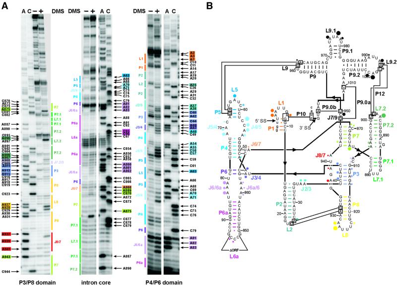
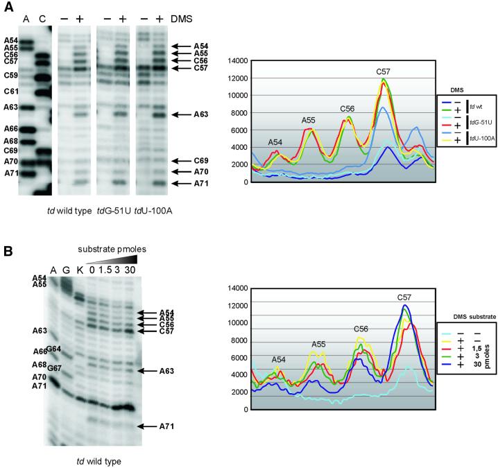
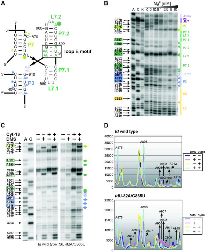
Group I introns consist of two major structural domains, the P4-P6 and P3-P9 domains, which assemble through interactions with peripheral extensions to fold into an active ribozyme. To assess group I intron folding in vivo, we probed the structure of td wild-type and mutant introns using dimethyl sulfate. The results suggest that the majority of the intron population is in the native state in accordance with the current structural model, which was refined to include two novel tertiary contacts. The importance of the loop E motif in the P7.1-P7.2 extension in assisting ribozyme folding was deduced from modeling and mutational analyses. Destabilization of stem P6 results in a deficiency in tertiary structure formation in both major domains, while weakening of stem P7 only interferes with folding of the P3-P9 domain. The different impact of mutations on the tertiary structure suggests that they interfere with folding at different stages. These results provide a first insight into the structure of folding intermediates and suggest a putative order of events in a hierarchical folding pathway in vivo.


Figures

None
Fig. 1. DMS modification of the td intron in vivo. (A) Intron residues accessible to DMS are displayed in these representative gels. Boxed nucleotides correspond to positions within the intron, which are modified by DMS. The P3-P8 domain of the intron core (left panel), the center of the intron core covering the P7 stem, as well as the P6-P6a element (middle panel) and the P4-P6 domain of the intron core and the stem–loops P1-P2 (right panel) are shown. The following color code is used: P1, orange; P2, turquoise; P3, blue-violet; P4 and P5, blue; P6 extension, purple; P7 extension, green; P8, yellow; and J8/7, red. (A) and (C) denote the sequencing lanes. (B) Summary of the td intron residues modified by DMS in vivo. Modified sites are indicated by dots. The size of the dots correlates with the relative modification intensities. The largest dot corresponds to the highest modification intensity [color code for the dots as in (A); modifications in P9 are represented by black dots]. This structure representation is based on that of Cech et al. (1994).
None
Fig. 2. Docking of the substrate helix to the P4-P5 A-rich asymmetric internal loop. Comparison of the accessibility of residues in J4/5 and J5/4 in vivo and in vitro. (A) The in vivo DMS modification pattern of the td wild-type intron and of td mutants harboring a nonsense codon in the upstream exon (tdG-51U, tdU-100A) is displayed in the left panel. Positions accessible to DMS are indicated by an arrow and base numbering. A and C are sequencing lanes. The corresponding phosphoimager quantification of these gel segments is shown in the right panel. (B) In vitro DMS modification of the ribozyme td WT-12 was performed in the presence of varying amounts of substrate (picomoles of substrate TDS4). A and G denote the sequencing lanes, and K represents the control lane, which derives from the reverse transcription of an RNA that was not treated with DMS. Quantification of the in vitro modification is shown in the right panel.
None
Fig. 3. The folding defect of splicing-deficient td intron mutants in vivo. (A) Changes within the modification pattern of the loop E motif and of the joining segment J8/7, which are due to a mutation in stem P6 (mutant tdU-82A/C865U), are displayed. The changes relative to the wild type are indicated as red triangles and the corresponding residue number is boxed in red. (B) Local changes within the td intron structure due to the mutation of the semi-conserved bulge in stem P7 (mutant tdC870U) are shown in the representative gels. Residues with an altered accessibility to DMS when compared with the wild-type intron are marked with blue triangles, and the corresponding nucleotide number is boxed in blue. (C) Quantification of the gel segments is shown for the td wild type and the mutant tdU-82A/C865U in the upper panel, as well as for the td wild type and the mutant tdC870U in the lower panel. (D) Summary of the changes in the DMS modification pattern occurring in the td intron mutants tdU-82A/C865U (red), tdC873U (yellow) and tdC870U (blue). The mutated residue is indicated by boxes labeled in the respective color. These changes are indicated relative to the wild-type td intron. Residues that are modified by DMS within these mutant introns, but whose modification intensity is not altered with respect to the wild-type construct, are not indicated (compare with Figure 1). Different dot sizes correlate to the relative increase in modification intensities compared with the wild type, whereas triangles indicate a reduced accessibility to DMS.
None
Fig. 4. Perturbation of the loop E motif. (A) The loop E motif of the P7.1-P7.2 extension of the td group I intron (boxed in black). The modified residues are labeled with dots whose size correlates to the relative modification intensities within the wild-type intron (color code as in Figure 1). (B) In vitro DMS modification of the ribozyme td WT-12 with increasing magnesium concentrations. Junction J6/6a to stem P8 is displayed. The increasing magnesium concentrations are indicated also. Boxed nucleotides on the left side of the gel correspond to positions within the intron whose DMS modification intensity changes with increasing magnesium concentration. The colored triangles represent a decrease in accessibility to DMS, whereas the green hexagon corresponds to an increased accessibility to DMS with increasing magnesium concentration. The color code is as in Figure 1. (C) The representative gels display the modification status of residues which are part of or close to the loop E motif in the context of the wild-type intron and of the mutant tdU-82A/C865U, in the absence and presence of the group I intron-specific splicing factor Cyt-18. The nucleotide numbering highlighted with colored boxes as well as the colored diamonds outline those residues whose accessibility was altered due to the mutation in stem P6. (D) Quantification of the modification intensities in the absence and presence of Cyt-18. The upper panel depicts the relative change in modification intensities of residues that are part of, or that are close to, the loop E motif within the wild-type intron, and in the lower panel within the mutant tdU-82A/C865U. Due to increased levels of td RNA in cells co-expressing Cyt-18, these differences were normalized (see Materials and methods).
None
Fig. 5. The refined 3D model of the td intron. (A) The global view of the intron points to the importance of the loop E motif (balls and sticks in red) both in the organization of the P7 extension and to the contacts between stem P3 and the P6/P6a A-rich internal loop (balls and sticks in orange). The remainder of the P7 extension is colored in green. The P4-P6 domain is marked in blue, while the P9 extension is shown in yellow. Stems P3 and P8 as well as junction J8/7 are labeled in purple. The helices P1 and P2 are outlined in gray. (B) The structure of the eukaryotic loop E motif was taken from Correll et al. (1999). It is described using the nomenclature proposed in Leontis and Westhof (2001): squares indicate that the base interacts via the Hoogsteen edge, triangles via the sugar edge and circles via the Watson–Crick edge. Open symbols are used for trans and full symbols for cis orientations of the glycosidic bonds relative to the hydrogen bonds. The sheared A·G pair involves hydrogen bonding between N6 of A and N3 of G, and between N7 of A and N2 of G. In the universally conserved trans Watson–Crick–Hoogsteen U·A pair, N3 of U interacts with N7 of A, and O2 of U contacts N6 of A. The N1 position of the bulged guanine residue is likely to be hydrogen-bonded to the phosphate oxygen of the cross strand A of the A·A pair. The symmetrical pairing between the trans Hoogsteen–Hoogsteen A·A pair involves hydrogen-bonding of the N7 and N6 positions of one A to the N6 and N7 positions of the other A. A typical example of contacts between a loop E motif and an RNA helix taken from the 23S rRNA of Haloarcula marismortui (Ban et al., 2000) is shown to the left of the one built into the td model. The underlined bases indicate those making the shallow groove contact the loop E motif. (C) The contacts between P3 and J6/6a, and a typical example from the 23S rRNA (Ban et al., 2000), are represented in a secondary-like structure diagram. In J6/6a, the dotted triangles on either side of the A·A pairs are meant to convey that the precise nature of the interactions cannot be fixed at this stage.

Similar articles

Cited by

References

    1. Ban N., Nissen,P., Hansen,J., Moore,P.B. and Steitz,T.A. (2000) The complete atomic structure of the large ribosomal subunit at 2.4 Å resolution. Science, 289, 905–920. - PubMed
    1. Belfort M., Chandry,P.S. and Pedersen-Lane,J. (1987) Genetic delineation of functional components of the group I intron in the phage T4 td gene. Cold Spring Harb. Symp. Quant. Biol., 52, 181–192. - PubMed
    1. Brion P., Michel,F., Schroeder,R. and Westhof,E. (1999a) Analysis of the cooperative thermal unfolding of the td intron of bacteriophage T4. Nucleic Acids Res., 27, 2494–2502. - PMC - PubMed
    1. Brion P., Schroeder,R., Michel,F. and Westhof,E. (1999b) Influence of specific mutations on the thermal stability of the td group I intron in vitro and on its splicing efficiency in vivo: a comparative study. RNA, 5, 947–958. - PMC - PubMed
    1. Caprara M.G., Lehnert,V., Lambowitz,A.M. and Westhof,E. (1996a) A tyrosyl-tRNA synthetase recognizes a conserved tRNA-like structural motif in the group I intron catalytic core. Cell, 87, 1135–1145. - PubMed

Publication types