. 2002 Oct;43(10):3181-9.

ECM-stimulated signaling and actin reorganization in embryonic corneal epithelia are Rho dependent

Affiliations

ECM-stimulated signaling and actin reorganization in embryonic corneal epithelia are Rho dependent

Wende R Reenstra et al. Invest Ophthalmol Vis Sci. 2002 Oct.

Abstract

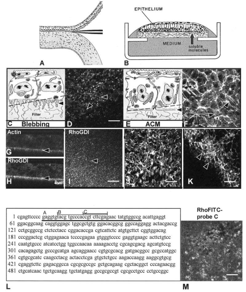
Purpose: The goal of this study was to investigate the role of the small guanosine triphosphatase (GTPase), Rho, in the corneal epithelial response to extracellular matrix (ECM) molecules. The avian corneal epithelial model was used to establish that Rho is required for actin reorganization and tyrosine phosphorylation of integrin-mediated signal pathway proteins.

Methods: Whole embryonic corneal epithelia were isolated without the basal lamina and either transfected with Rho-specific antisense oligonucleotides or treated with Clostridium botulinum C3 exoenzyme and then stimulated with fibronectin (FN) or collagen (COL). The epithelia were evaluated for actin reorganization and protein production including Rho protein levels and tyrosine phosphorylation with Western blot analysis.

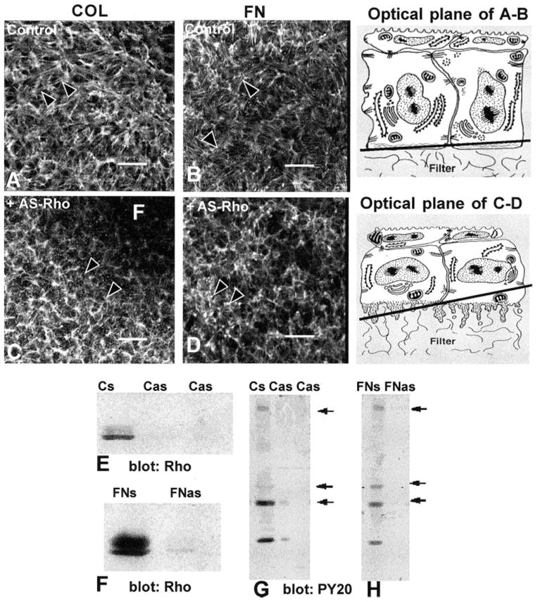
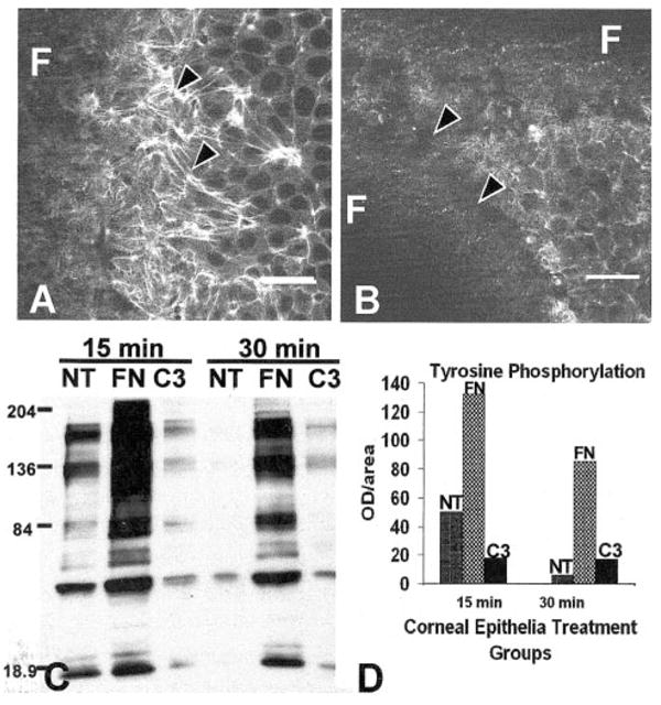
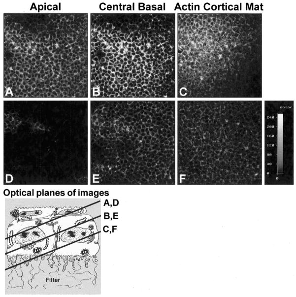
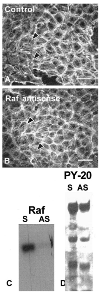
Results: After an overnight transient transfection with antisense oligonucleotides, Rho protein levels were decreased more than 80%, and tyrosine phosphorylation of all integrin-mediated signal transduction proteins was decreased compared with control epithelia. Intracellular Rho distribution did not change in the presence of antisense oligonucleotides; however, the amount of immunolabeled Rho decreased. Disrupting the signaling cascade with Rho antisense also blocked FN- and COL-stimulated actin cortical mat reformation. C. botulinum C3 exoenzyme, a pharmacologic agent that specifically causes adenosine diphosphate (ADP) ribosylation and inactivation of Rho, also blocked actin reorganization and tyrosine phosphorylation. In contrast, decreasing Raf protein levels did not change FN-mediated actin reorganization or tyrosine phosphorylation.

Conclusions: Decreasing Rho protein or blocking its function inhibited ECM-stimulated actin reorganization and signal transduction, as measured by tyrosine phosphorylation.


Figures

Figure 1
Figure 1
Schematic drawing of isolation of corneal epithelium (A), culture of the cells (B), and confocal images (D, FK), human Rho A sequence (L), antisense probes (L, A–C) and confocal image of FITC-labeled antisense oligonucleotide probe (M). Epithelia were isolated as a sheet of tissue (A), placed on a polycarbonate filter basal-side down, and cultured at the air–medium interface (B). Epithelia isolated without the basal lamina extended basal cellular processes termed blebs (C, arrowhead). The blebs contain F-actin, which appeared as punctate spots in single optical sections (D, arrowheads) taken through the basal area illustrated in the schematic drawing (C, dashed line). Immunohistochemical localization of RhoGDI demonstrated that this protein was enriched in the protruding blebs from tissues isolated without the basal lamina (H, I). Tissue cultured with soluble ECM molecules reorganized the basal actin into an ACM (E, arrowhead). The bundles of F-actin viewed in a single optical section from epithelia stimulated with COL for 2 hours (F, arrowheads) appeared to align from cell to cell, across the field. The epithelium stimulated with bombesin (J) or LPA (K) reformed the ACM after 30 minutes. The upper right corner (K) was in the optical section through the basal cell nuclei, and the individual cell size was visible in the honeycomb pattern of the lateral cell membrane actin distribution. Corneal epithelial tissues were cultured in control media, transfected with a 40- (L-A), 20- (L-B), or 10-mer (L-C) oligonucleotide directed against the 5′ region of human Rho mRNA (5–10 ng per 20 epithelia) for 24 hours in the presence of liposomes. The largest probe (L-A) was used first, and then the shorter probes were designed. A single optical section through the basal cells demonstrates that the oligonucleotide (FITC-labeled 10-mer) was evenly transfected throughout the tissue (M). Scale bar: (D, IK, M) 20 μm; (G, H) 8 μm.
Figure 2
Figure 2
Corneal epithelia transiently transfected (24 hours) with antisense Rho oligonucleotides (10-mer, Fig. 1L-C) were stimulated with FN or COL for 2 hours and stained with fluorescent phalloidin. All experiments and procedures were repeated at least three times with similar results. Single confocal optical sections through basal cells, as illustrated in the tissue schematic, show the distribution of the F-actin in epithelia stimulated with either COL or FN in the control tissues (A, B). Areas that did not contain actin staining (dark) were optical sections through the supporting filter (F). The tissues were not flat; therefore, a single optical section may contain the filter (F) and basal cell cytoplasm (C). In tissues transiently transfected with Rho antisense (C, D), the epithelia had decreased actin in the basal compartment and did not form an ACM after stimulation with COL or FN. The optical sections were taken at the base of the epithelia, as illustrated in the schematic at various tangential planes. Epithelia harvested and analyzed with Western blot analysis were probed with anti-Rho (E, F). The sense control (FNs or Cs) had a 21-kDa protein that migrated as a doublet. In contrast, epithelia transfected with antisense Rho had 80% less Rho protein (E, F; Cas, FNas). To determine whether Rho protein affects other signal transduction proteins, the same Western blot analysis used in (E) and (F) were stripped and reprobed with anti-phosphotyrosine (G, H). In control tissues (Cs, FNs), tyrosine-phosphorylated proteins at molecular weights of approximately 190, 68, 42, and 20 kDa were detectable (arrows), whereas in the tissue treated with antisense Rho oligonucleotides (Cas, FNas), these proteins were significantly decreased. Scale bar, 20 μm.
Figure 3
Figure 3
Corneal epithelia treated with exoenzyme C3 (3 μg/mL) did not reorganize the ACM (B) compared with the control (A) and decreased tyrosine phosphorylation (C, D) compared with FN-stimulated epithelia. The total optical density from this sample immunoblot using the whole lane demonstrated that C3 decreased tyrosine phosphorylation 8- to 10-fold (D). All experiments and procedures were repeated at least three times with similar results. Single confocal optical sections through basal cells, as illustrated in the tissue schematic (Fig. 2), show the distribution of the F-actin in epithelia stimulated with FN in the control tissues (A). Areas that did not contain actin staining (dark) were optical sections through the supporting filter (F). The tissues were not flat; therefore, a single optical section may contain the filter (F) and basal cell cytoplasm. Scale bar, 20 μm.
Figure 4
Figure 4
Corneal epithelial cells were transfected with sense or antisense oligonucleotides directed toward Rho, immunolabeled with anti-Rho antibody, and analyzed with by CLSM, to determine intracellular distribution and transfection efficiency. The pinhole, voltage, and offset were the set at the same levels on both images. The intensity wedge indicates the range of detected light intensity from 0 (black) to 255 (white). Rho in control sense-transfected epithelia had a honeycomb distribution, similar to other membrane-associated proteins (AC). The protein was less prominent in the periderm cells but was evenly distributed throughout the basal cells in a membrane-bound pattern (AC). In contrast, epithelia transfected with antisense Rho had significantly less immunolabeled protein, indicating that the transfection decreased the amount of Rho protein evenly throughout the epithelium (DF). The intensity wedge indicates the relative intensity of the pixels in each image. The schematic drawing indicates the plane of the optical sections. All images are single optical sections through representative tissues from three experiments.
Figure 5
Figure 5
Corneal epithelia transiently transfected (24 hours) with antisense Raf oligonucleotides (10-mer directed to the 5′ region) and stimulated with FN for 2 hours had a normal actin distribution (A, B) and tyrosine phosphorylation pattern (D). All experiments were repeated at least three times with similar results. Single confocal optical sections through basal cells (as illustrated in the schematic in Fig. 2) demonstrated the F-actin distribution in epithelia stimulated with FN in the presence of Raf sense (A) or antisense (B) oligonucleotides formed an ACM. Epithelia harvested and analyzed with Western blot analysis probed with anti-Raf (C). The sense control had protein that migrated at the appropriate molecular weight. In contrast, epithelia transfected with antisense Raf had less Raf protein (C). To determine whether Raf protein affected other signal transduction proteins, the same Western blot shown in (C) was stripped and reprobed with anti-phosphotyrosine. Nearly all the phosphoproteins found in the sense control were also present in the antisense-treated tissue (D).

Similar articles

Cited by

References

    1. Zieske JD. Perpetuation of stem cells in the eye. Eye. 1994;8:163 – 169. - PubMed
    1. Gipson I, Inatomi F. Extracellular matrix and growth factors in corneal wound healing. Curr Opin Ophthalmol. 1995;6:3 – 10. - PubMed
    1. Nishida T, Tanaka T. Extracellular matrix and growth factors in corneal wounding. Curr Opin Ophthalmol. 1996;7:2 – 11. - PubMed
    1. Sundarraj N, Kinchington PR, Wessel H, et al. A Rho-associated protein kinase: differentially distributed in limbal and corneal epithelia. Invest Ophthalmol Vis Sci. 1998;39:1266 – 1272. - PubMed
    1. Anderson SC, Sundarraj N. Regulation of a Rho-associated kinase expression during the corneal epithelial cell cycle. Invest Ophthalmol Vis Sci. 2001;42:933 – 940. - PubMed

Publication types