. 2002 Sep 30;158(7):1263-75.
doi: 10.1083/jcb.200203043.

T cell receptor ligation induces the formation of dynamically regulated signaling assemblies

Affiliations

T cell receptor ligation induces the formation of dynamically regulated signaling assemblies

Stephen C Bunnell et al. J Cell Biol. .

Abstract

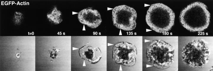
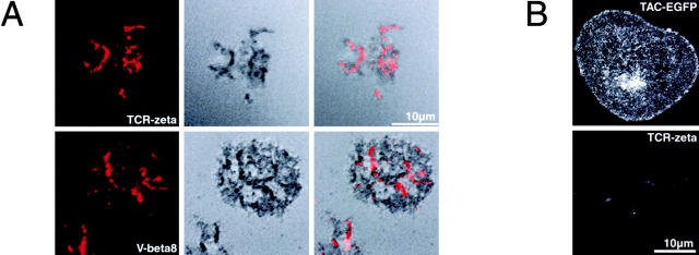
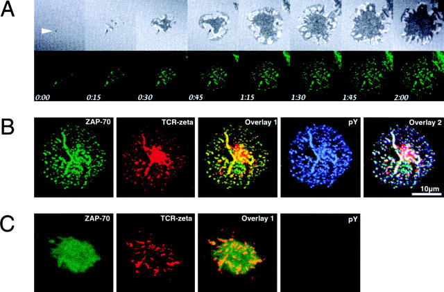
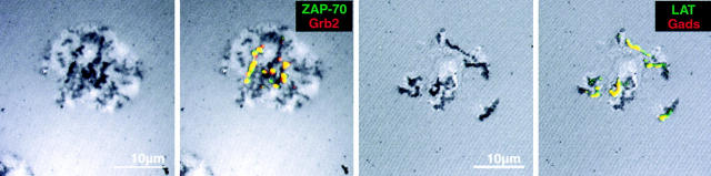
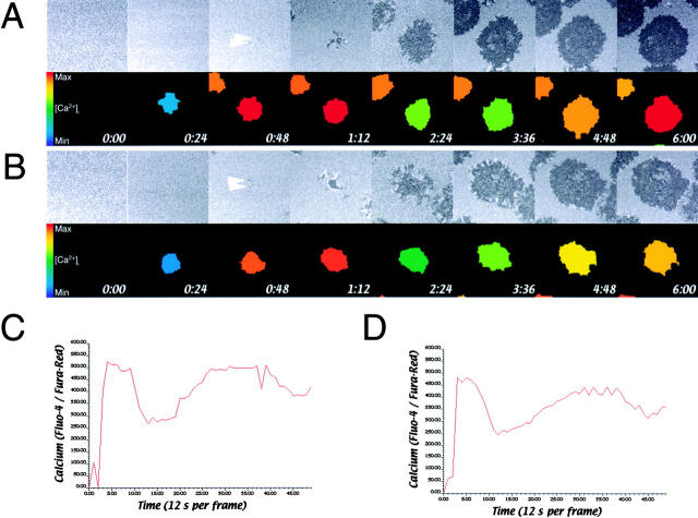
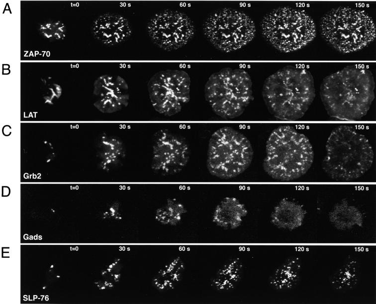
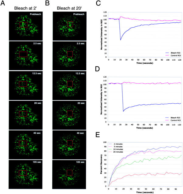
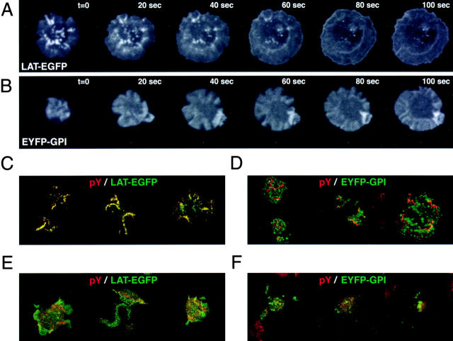
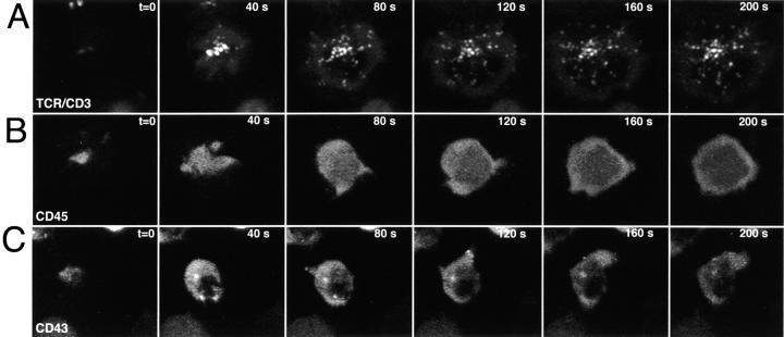
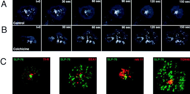
Tcell antigen receptor (TCR) ligation initiates tyrosine kinase activation, signaling complex assembly, and immune synapse formation. Here, we studied the kinetics and mechanics of signaling complex formation in live Jurkat leukemic T cells using signaling proteins fluorescently tagged with variants of enhanced GFP (EGFP). Within seconds of contacting coverslips coated with stimulatory antibodies, T cells developed small, dynamically regulated clusters which were enriched in the TCR, phosphotyrosine, ZAP-70, LAT, Grb2, Gads, and SLP-76, excluded the lipid raft marker enhanced yellow fluorescent protein-GPI, and were competent to induce calcium elevations. LAT, Grb2, and Gads were transiently associated with the TCR. Although ZAP-70-containing clusters persisted for more than 20 min, photobleaching studies revealed that ZAP-70 continuously dissociated from and returned to these complexes. Strikingly, SLP-76 translocated to a perinuclear structure after clustering with the TCR. Our results emphasize the dynamically changing composition of signaling complexes and indicate that these complexes can form within seconds of TCR engagement, in the absence of either lipid raft aggregation or the formation of a central TCR-rich cluster.


Figures

Figure 1.
Figure 1.
The rearrangement of the actin cytoskeleton is coupled to contact formation. Jurkat T cells expressing EGFP-actin were plated on stimulatory coverslips and imaged using a Zeiss LSM 410 confocal system. EGFP-actin and IRM images of the contact were collected every 45 s. Arrowheads emphasize the correspondence between the actin rich structures (top) and the developing contacts (bottom).
Figure 2.
Figure 2.
The TCR is specifically clustered within tight contacts. (A) Jurkat cells were either plated on HIT3a-coated coverslips, fixed after 1 min and stained for TCRζ (top), or plated on UCHT1-coated coverslips, fixed after 2 min and stained for Vβ8 (bottom). Cells were then permeabilized and stained for TCR components (left). Contacts between stained T cells and the coverslip were also imaged by IRM, where tight contacts appear darker than loose contacts (middle). Overlays reveal that receptor engagement occurs preferentially in the zones of tightest contact (right). (B) TAC–EGFP-expressing Jurkat T cells were plated on coverslips and fixed after 5 min. In the top panel, cells were plated on stimulatory HIT3a-coated coverslips and imaged for EGFP. In contrast, in the bottom panel, cells were plated on anti–TAC-coated coverslips and stained for TCRζ. No clustering is apparent in either sample.
Figure 3.
Figure 3.
ZAP-70 is specifically recruited to activated TCRs in tight contacts. (A) Jurkat T cells expressing ZAP-70–EGFP were plated on coverslips. ZAP-70–EGFP and IRM images were collected every 15 s using the Zeiss LSM 410. The white arrow marks the earliest observed signaling cluster and contact point. (B) Jurkat T cells expressing ZAP-70–EGFP were plated on stimulatory coverslips, fixed after 5 min, and stained for both TCRζ and phosphotyrosine. ZAP-70–EGFP, TCRζ, and phosphotyrosine were pseudocolored green, red, and blue, respectively. The TCRζ staining pattern shows a large cluster in the center of the contact; this strong signal results from the staining of a large, perinuclear pool of TCRζ. (C) Cells were processed and imaged as in B after pretreatment and stimulation in the presence of 10 μM PP2.
Figure 4.
Figure 4.
Signal transducing complexes form at tight contacts. Jurkat T cells stably expressing either ZAP-EGFP or LAT-EGFP (green) were plated on stimulatory coverslips, fixed after 2 min, permeabilized, and stained for either Grb2 or Gads (red). IRM images (left) and fluorescent overlays on the IRM images (right) are shown. Note the strong correspondence between tight contacts and clusters of signaling proteins.
Figure 5.
Figure 5.
Small contacts are sufficient to initiate calcium influxes. (A and B) Jurkat T cells loaded with the calcium responsive dyes Fluo-4 and Fura-Red were plated on coverslips and dynamically imaged for calcium content and T cell-coverslip contact. In the bottom panels the bodies of responding T cells are pseudocolored to indicate their calcium content. The pseudocolored calcium scale is inset in the leftmost panels. White arrows mark the earliest observed contact points. (C and D) Traces of calcium over time for the two cells shown in A and B. The time axis is marked in frames; one frame corresponds to 12 s. These traces are representative of those obtained from >30 cells imaged in three separate experiments.
Figure 6.
Figure 6.
Signaling adaptors are dynamically redistributed throughout contact formation. (A–E) Live Jurkat E6.1 cells expressing EGFP- or EYFP-chimeras were plated on stimulatory coverslips and observed dynamically using the Ultraview spinning wheel confocal system. Seven-image, 3.5 μm deep Z stacks were collected every 10 s for ZAP-70, every 15 s for the remaining proteins.
Figure 7.
Figure 7.
Photobleaching reveals that ZAP-70 equilibrates between signaling complexes. (A and B) ZAP-70–EGFP-expressing Jurkat T cells were plated on coverslips and allowed to develop contacts for either 2 or 20 min. Contacts were imaged for 20 s, photobleached within a specific region of interest (ROI, red boxes), and monitored for the recovery of fluorescence in the bleached ROI. Images were obtained every 2.5 s. (C) Mean fluorescence recovery traces from cells plated for 2 min before photobleaching (n = 6). (D) Mean fluorescence recovery traces from cells plated for 20 min before photobleaching (n = 6). (E) The extent of fluorescence recovery in bleached ROIs as a function of the age of the observed contacts at the time of photobleaching. In each case, half-maximal recovery is observed within 7.5 to 10 s.
Figure 8.
Figure 8.
LAT is selectively retained in signaling complexes. (A and B) Jurkat cells expressing either LAT-EGFP or EYFP-GPI were plated on stimulatory coverslips and imaged using the Ultraview system. Five-image, 2.5 μm deep Z stacks were collected every 20 s. The bright spot observed in the EYFP-GPI–expressing cells corresponds to a membrane fold, not a cluster. Similar structures are also occasionally observed with Tac-EGFP, a nonraft membrane protein. (C and D) Jurkat cells expressing either LAT-EGFP or EYFP-GPI (green) were plated on stimulatory coverslips, fixed after 2 min, and stained for phosphotyrosine (red). (E and F) Jurkat cells expressing either LAT-EGFP or EYFP-GPI were treated with 10 μM PP2, and then plated and imaged in C and D.
Figure 9.
Figure 9.
SLP-76 clustering is selectively induced by TCR ligation. (A and C) SLP-76–EYFP expressing J14 cells were plated on antibody-coated coverslips and imaged using the Ultraview system. Five-image, 2.5 μm deep Z stacks were collected every 10 s. The plate-bound antibodies were specific for (A) CD3ɛ, (B) CD45, or (C) CD43.
Figure 10.
Figure 10.
SLP-76–containing clusters move on microtubules to an undefined perinuclear compartment. (A and B) Jurkat J14 cells expressing SLP-76–EYFP were plated on stimulatory coverslips and imaged as in Fig. 9 after 1 h pretreatment with either DMSO carrier (A) or 100 μM colchicine (B). Drugs were also present throughout imaging. (C) The colocalization of SLP-76–EYFP with various intracellular markers was tested in both live and fixed cells. Jurkat J14 cells expressing SLP-EGFP (green) were serum starved for 40 min, incubated with 100 μM transferrin-Alexa 594 (red), plated on stimulatory coverslips and imaged using the Ultraview system. Every 20 s, individual red and green image stacks were collected. Each image stack is five images (2.5 μm) deep. The image shown here was collected 2 min into the spreading process (panel 1). Jurkat J14 cells expressing SLP-76–EYFP (green) were also plated on stimulatory coverslips, fixed after 10 min, and stained for intracellular markers (red): (panel 2) EEA1; (panel 3) rab11; or (panel 4) TGN46.

References

    1. Allenspach, E.J., P. Cullinan, J. Tong, Q. Tang, A.G. Tesciuba, J.L. Cannon, S.M. Takahashi, R. Morgan, J.K. Burkhardt, and A.I. Sperling. 2001. ERM-dependent movement of CD43 defines a novel protein complex distal to the immunological synapse. Immunity. 15:739–750. - PubMed
    1. Anton van der Merwe, P., S.J. Davis, A.S. Shaw, and M.L. Dustin. 2000. Cytoskeletal polarization and redistribution of cell-surface molecules during T cell antigen recognition. Semin. Immunol. 12:5–21. - PubMed
    1. Balamuth, F., D. Leitenberg, J. Unternaehrer, I. Mellman, and K. Bottomly. 2001. Distinct patterns of membrane microdomain partitioning in Th1 and Th2 cells. Immunity. 15:729–738. - PubMed
    1. Boniface, J.J., J.D. Rabinowitz, C. Wulfing, J. Hampl, Z. Reich, J.D. Altman, R.M. Kantor, C. Beeson, H.M. McConnell, and M.M. Davis. 1998. Initiation of signal transduction through the T cell receptor requires the multivalent engagement of peptide/MHC ligands. Immunity. 9:459–466. - PubMed
    1. Brdicka, T., D. Pavlistova, A. Leo, E. Bruyns, V. Korinek, P. Angelisova, J. Scherer, A. Shevchenko, I. Hilgert, J. Cerny, et al. 2000. Phosphoprotein associated with glycosphingolipid-enriched microdomains (PAG), a novel ubiquitously expressed transmembrane adaptor protein, binds the protein tyrosine kinase csk and is involved in regulation of T cell activation. J. Exp. Med. 191:1591–1604. - PMC - PubMed

Publication types

MeSH terms