. 2002 Sep 30;158(7):1277-85.
doi: 10.1083/jcb.200208083.

CD40, an extracellular receptor for binding and uptake of Hsp70-peptide complexes

Affiliations

CD40, an extracellular receptor for binding and uptake of Hsp70-peptide complexes

Thalia Becker et al. J Cell Biol. .

Abstract

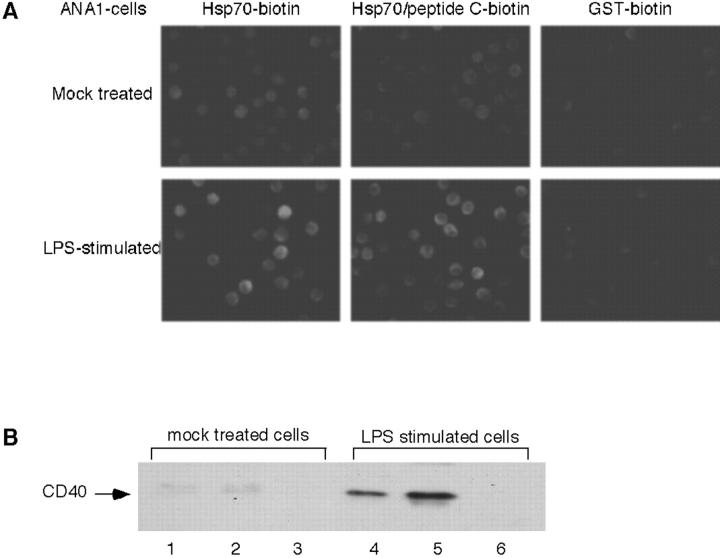
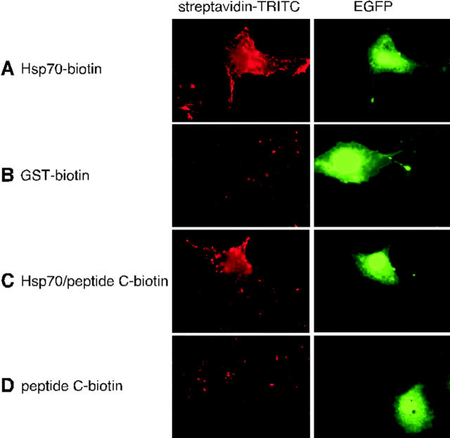
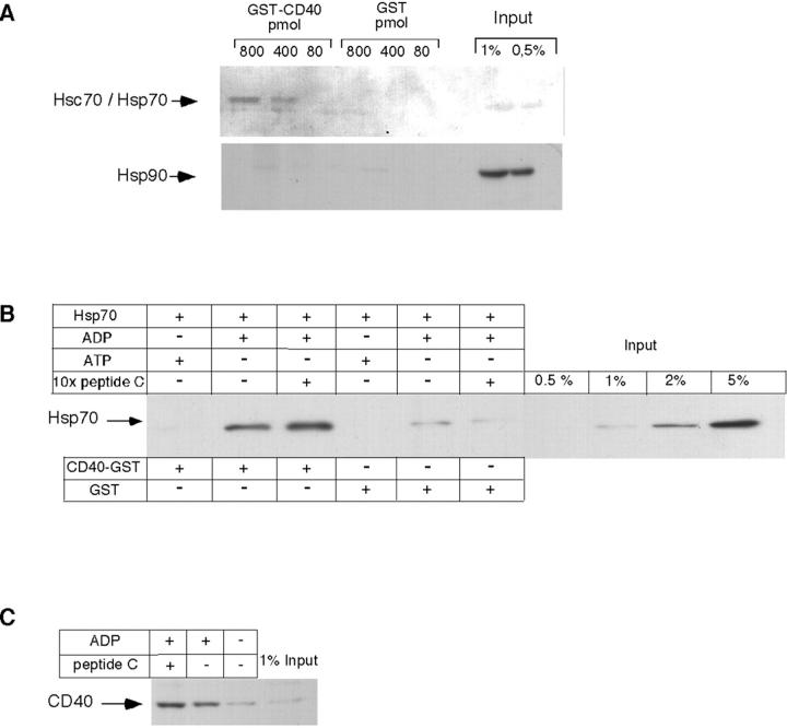
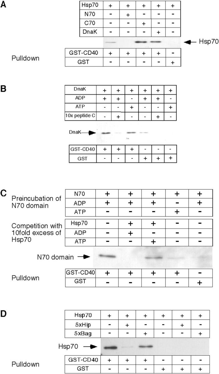
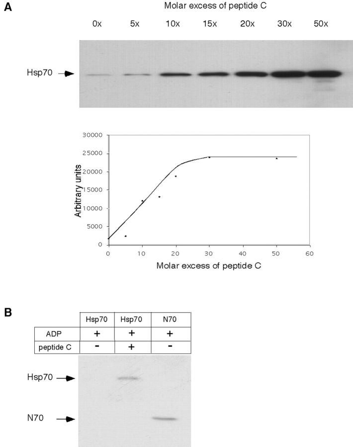
Tumor and viral antigens elicit a potent immune response by heat shock protein-dependent uptake of antigenic peptide with subsequent presentation by MHC I. Receptors on antigen-presenting cells that specifically bind and internalize a heat shock protein-peptide complex have not yet been identified. Here, we show that cells expressing CD40, a cell surface protein crucial for B cell function and autoimmunity, specifically bind and internalize human Hsp70 with bound peptide. Binding of Hsp70-peptide complex to the exoplasmic domain of CD40 is mediated by the NH(2)-terminal nucleotide-binding domain of Hsp70 in its ADP state. The Hsp70 cochaperone Hip, but not the bacterial Hsp70 homologue DnaK, competes formation of the Hsp70-CD40 complex. Binding of Hsp70-ADP to CD40 is strongly increased in the presence of Hsp70 peptide substrate, and induces signaling via p38. We suggest that CD40 is a cochaperone-like receptor mediating the uptake of exogenous Hsp70-peptide complexes by macrophages and dendritic cells.


Figures

Figure 1.
Figure 1.
LPS treatment of ANA-1 cells stimulates binding of Hsp70 and induces expression of CD40. Cells were incubated with LPS or mock-treated as outlined in Materials and methods. (A) Cells were incubated either with biotinylated Hsp70, Hsp70 loaded with biotinylated peptide C, or with biotinylated GST as a control. After 30 min at 4°C, cells were washed, incubated with TRITC-labeled streptavidin, washed again, and processed for fluorescence microscopy. (top) mock-treated ANA-1 cells; (bottom) ANA-1 cells after treatment with LPS. Note that Hsp70 carries on average five biotins, whereas peptide C contains a single biotin. (B) Cells were harvested, lysed, and centrifuged to obtain a total membrane pellet. Identical protein amounts of the samples were analyzed by immunoblotting with an antibody directed against CD40. (Lanes 1 and 4) total cell lysates; (lanes 2 and 5) membrane fractions; (lanes 3 and 6) supernatants.
Figure 2.
Figure 2.
Transfection with human CD40 cDNA renders Cos-7 cells active in binding human Hsp70. Cos-7 cells were transiently transfected with a fusion construct that contained human CD40 cDNA, followed by an internal ribosomal entry site and the cDNA for EGFP, giving rise to green fluorescence of transfected cells. Cells were incubated for 30 min at 0°C with biotinylated Hsp70 (A), biotinylated GST as a control (B), Hsp70–peptide complex containing biotinylated peptide C (C), or with biotinylated peptide C alone (D). Thereafter, cells were washed and incubated with TRITC-streptavidin and processed for fluorescence microscopy as described in Fig. 1. Left-hand panels show streptavidin fluorescence and right-hand panels show EGFP fluorescence.
Figure 3.
Figure 3.
Binding of Hsp70 to CD40 is direct and depends on ADP. (A) HeLa cell lysates were incubated with GST (control) or GST-CD40 in the amounts indicated. Thereafter, samples were affinity-purified on glutathione-sepharose as outlined in Materials and methods, subjected to SDS-PAGE, and analyzed by immunoblotting with antibodies directed against Hsc70 and Hsp70 (top lanes), and against Hsp90 (bottom lanes). (B) Human recombinant His6-tagged Hsp70 was incubated with ATP, ADP, or an excess of peptide C, followed by addition of CD40-GST or GST alone. After affinity purification, samples were analyzed by immunoblotting as described in A, using antibodies directed against the His6 tag. (C) Recombinant human His6-tagged Hsp70 was incubated with peptide C, either in the presence or absence of ADP. The reactions were then incubated with ANA-1 cell lysates as described in Materials and methods. Protein bound to Hsp70 was analyzed by affinity purification on Ni-NTA agarose and immunoblotting with antibodies directed against CD40. Input reflects the amount of protein subjected to affinity purification.
Figure 4.
Figure 4.
Hsp70 binding to CD40 is mediated by the NH2-terminal ATPase domain and is competed by Hip. (A) Human His6-tagged Hsp70, its NH2- or COOH-terminal domains, or recombinant bacterial DnaK was incubated either with GST-CD40 or with GST. (B) Recombinant DnaK was incubated with ADP, ATP, or an excess of peptide C, followed by addition of GST-CD40 or GST alone. (C) His6-tagged N70 was incubated in the presence of ADP or ATP, followed by incubation with a 10-fold molar excess of Hsp70 in the presence of ADP or ATP. (D) Recombinant human His6-tagged Hsp70 protein was incubated with a fivefold molar excess of either recombinant Hip protein or Bag-1, and with GST-CD40 or GST as a control. Bound protein was analyzed after affinity purification on glutathione-sepharose by immunoblotting with an antibody directed against the NH2-terminal His6 tags, or with an antibody directed against DnaK.
Figure 5.
Figure 5.
Peptide substrate stimulates Hsp70 binding to CD40. (A) His6-tagged Hsp70 was incubated with increasing concentrations of peptide C as indicated, and binding to GST-CD40 was analyzed by immunoblotting with antibodies directed against the His6-tag. The bottom panel is a quantitation of the data. (B) Equivalent concentrations (3 μM) of His6-tagged Hsp70 (in the presence of 2 mol ADP and in the absence or presence of a 30-fold molar excess of peptide C) or His6-tagged N70 were incubated with GST-CD40, and bound protein was analyzed as described for panel A.
Figure 6.
Figure 6.
Binding of Hsp70 complex to CD40-expressing HEK293T cells induces signaling via p38 and causes peptide uptake. (A) HEK293T cells, stably transfected with cDNA encoding human CD40 or the unrelated membrane protein (MCAT) were incubated for 20 min at 37°C with CD40L or with Hsp70, N70, C70, or bacterial DnaK in the presence of peptide C and ADP or AMPPNP, or buffer alone. Thereafter, cells were washed, solubilized in SDS-sample buffer, and analyzed by immunoblotting with antibodies directed against phosphorylated (active) p38. Blots were also developed with an antibody against tubulin in order to control for equal loading. (B) Cells were incubated at 0°C for 30 min with recombinant human Hsp70, N70, or C70, all in the presence FITC-labeled peptide, or with the FITC-labeled peptide alone. Thereafter, cells were washed, incubated for 15 min at 37°C, and processed for fluorescence microscopy. (1) Hsp70, (2) FITC-labeled peptide alone, (3) C70, and (4) N70. Only cells stably transfected with CD40 are shown. Cell boundaries are emphasized with broken lines. MCAT-expressing control cells did not show fluorescence above background.
Figure 7.
Figure 7.
Model for the release of Hsp70–peptide complex from a necrotic tumor cell, followed by binding and uptake by an APC. (1) Necrosis leads to cell swelling and a concomitant drop in the concentration ratio of ATP/ADP. Intracellular Hsp70 is bound to unfolded protein or peptide. (2) Release of Hsp70–peptide complexes upon cell lysis. At the low nucleotide concentrations prevailing in the extracellular space, the half-life of the Hsp70-ADP state determines the stability of the Hsp70–peptide complex. (3) Hsp70–peptide complex binds to the surface of an APC. (4) Cell surface binding is mediated by the extracytoplasmic domain of CD40. (5) Hsp70-peptide binding alters the trimeric structure of CD40 (Chan et al., 2000), followed by activation of signaling via p38. (6), Hsp70–peptide complex is internalized by receptor-mediated endocytosis.

Similar articles

Cited by

References

    1. Arnold-Schild, D., D. Hanau, D. Spehner, C. Schmid, H.G. Rammensee, H. de la Salle, and H. Schild. 1999. Cutting edge: receptor-mediated endocytosis of heat shock proteins by professional antigen-presenting cells. J. Immunol. 162:3757–3760. - PubMed
    1. Asea, A., M. Rehli, E. Kabingu, J.A. Boch, O. Bare, P.E. Auron, M.A. Stevenson, and S.K. Calderwood. 2002. Novel signal transduction pathway utilized by extracellular HSP70: role of toll-like receptor (TLR) 2 and TLR4. J. Biol. Chem. 277:15028–15034. - PubMed
    1. Basu, S., and P.K. Srivastava. 1999. Calreticulin, a peptide-binding chaperone of the endoplasmic reticulum, elicits tumor- and peptide-specific immunity. J. Exp. Med. 189:797–802. - PMC - PubMed
    1. Basu, S., R.J. Binder, R. Suto, K.M. Anderson, and P.K. Srivastava. 2000. Necrotic but not apoptotic cell death releases heat shock proteins, which deliver a partial maturation signal to dendritic cells and activate the NF-κB pathway. Int. Immunol. 12:1539–1546. - PubMed
    1. Berwin, B., and C.V. Nicchitta. 2001. To find the road traveled to tumor immunity: the trafficking itineraries of molecular chaperones in antigen-presenting cells. Traffic. 2:690–697. - PubMed

MeSH terms