. 2002 Sep 30;158(7):1287-97.
doi: 10.1083/jcb.200207008.

P21-activated kinase 4 interacts with integrin alpha v beta 5 and regulates alpha v beta 5-mediated cell migration

Affiliations

P21-activated kinase 4 interacts with integrin alpha v beta 5 and regulates alpha v beta 5-mediated cell migration

Hongquan Zhang et al. J Cell Biol. .

Abstract

p21-activated kinase 1 (PAK1) can affect cell migration (Price et al., 1998; del Pozo et al., 2000) and modulate myosin light chain kinase and LIM kinase, which are components of the cellular motility machinery (Edwards, D.C., L.C. Sanders, G.M. Bokoch, and G.N. Gill. 1999. Nature Cell Biol. 1:253-259; Sanders, L.C., F. Matsumura, G.M. Bokoch, and P. de Lanerolle. 1999. SCIENCE: 283:2083-2085). We here present a novel cell motility pathway by demonstrating that PAK4 directly interacts with an integrin intracellular domain and regulates carcinoma cell motility in an integrin-specific manner. Yeast two-hybrid screening identified PAK4 binding to the cytoplasmic domain of the integrin beta 5 subunit, an association that was also found in mammalian cells between endogenous PAK4 and integrin alpha v beta 5. Furthermore, we mapped the PAK4 binding to the membrane-proximal region of integrin beta 5, and identified an integrin-binding domain at aa 505-530 in the COOH terminus of PAK4. Importantly, engagement of integrin alpha v beta 5 by cell attachment to vitronectin led to a redistribution of PAK4 from the cytosol to dynamic lamellipodial structures where PAK4 colocalized with integrin alpha v beta 5. Functionally, PAK4 induced integrin alpha v beta 5-mediated, but not beta1-mediated, human breast carcinoma cell migration, while no changes in integrin cell surface expression levels were observed. In conclusion, our results demonstrate that PAK4 interacts with integrin alpha v beta 5 and selectively promotes integrin alpha v beta 5-mediated cell migration.


Figures

Figure 1.
Figure 1.
PAK4 interacts with the integrin β5 subunit. (A) Amino acid sequence comparison of mPAK4 KD, found by the yeast two-hybrid screening with hPAK4, Drosophila MBT (MBT), and hPAK1 KDs. Dashes stand for identical amino acids and the variations of amino acids are indicated by letters in hPAK4, MBT, and hPAK1 sequences. The mPAK4 KD is numbered according to that of hPAK4 (Abo et al., 1998). (B) PAK4 association with integrin β5 in GST pull-down assays. GST–PAK4 KD fusion protein pulled down endogenous integrin β5 in cell lysates (top panel). GST–β5 cytoplasmic domain fusion protein pulled down overexpressed HA–PAK4 (bottom). The positive controls represent Western blot of the input lysates for marking the size and showing the presence of integrin β5 and PAK4. (C) PAK4 association with integrins in living cells. (C, top) Anti–integrin β3 mAb AP3 and anti–integrin αvβ5 mAb P1F6 coimmunoprecipitation of PAK4 in COS-7 cells expressing HA–PAK4. (C, middle and bottom) An anti-HA mAb coimmunoprecipitated HA–PAK4 with both αv and β5 integrin subunits. The positive controls represent Western blot of the input lysates for marking the size and showing the presence of integrins and PAK4. (D) Association of endogenous PAK4 with endogenous integrin αvβ5 in MCF-7 cells analyzed by IP. IP using rabbit IgG or α-Rab1 are negative controls.
Figure 1.
Figure 1.
PAK4 interacts with the integrin β5 subunit. (A) Amino acid sequence comparison of mPAK4 KD, found by the yeast two-hybrid screening with hPAK4, Drosophila MBT (MBT), and hPAK1 KDs. Dashes stand for identical amino acids and the variations of amino acids are indicated by letters in hPAK4, MBT, and hPAK1 sequences. The mPAK4 KD is numbered according to that of hPAK4 (Abo et al., 1998). (B) PAK4 association with integrin β5 in GST pull-down assays. GST–PAK4 KD fusion protein pulled down endogenous integrin β5 in cell lysates (top panel). GST–β5 cytoplasmic domain fusion protein pulled down overexpressed HA–PAK4 (bottom). The positive controls represent Western blot of the input lysates for marking the size and showing the presence of integrin β5 and PAK4. (C) PAK4 association with integrins in living cells. (C, top) Anti–integrin β3 mAb AP3 and anti–integrin αvβ5 mAb P1F6 coimmunoprecipitation of PAK4 in COS-7 cells expressing HA–PAK4. (C, middle and bottom) An anti-HA mAb coimmunoprecipitated HA–PAK4 with both αv and β5 integrin subunits. The positive controls represent Western blot of the input lysates for marking the size and showing the presence of integrins and PAK4. (D) Association of endogenous PAK4 with endogenous integrin αvβ5 in MCF-7 cells analyzed by IP. IP using rabbit IgG or α-Rab1 are negative controls.
Figure 2.
Figure 2.
Mapping of the PAK4 binding region within the integrin β5 cytoplasmic domain. (A) Mapping of the PAK4-binding region in the integrin β5 cytoplasmic domain. Various regions of the integrin β5 cytoplasmic domain were cloned into the bait vector pEG202 and then mated with PAK4 KD in the prey vector in a yeast two-hybrid assay. The PAK4 binding region was mapped to aa 759–767 of the β5 cytoplasmic domain. (B) Association of endogenous PAK4 with the membrane-proximal region within integrin β5 cytoplasmic domain was examined by a GST pull-down assay, including GST fused to a β5 deletion mutant lacking the PAK4-binding region mapped by yeast two-hybrid analysis. (C) Sequence comparison of the PAK4-binding region of integrin β5 with other integrin cytoplasmic domains (top). The Rack1- (Liliental and Chang, 1998) and PAK4-binding regions within integrin β5 are indicated (bottom).
Figure 3.
Figure 3.
Mapping of the integrin β5–binding region within the PAK4 KD. (A) Various regions of the PAK4 KD were cloned into the prey vector pJG4-5 and then mated in a yeast two-hybrid assay with the integrin β5 cytoplasmic domain in the bait vector. Amino acids 505–530 in the PAK4 KD were identified as the responsible region for interaction with the integrin β5 cytoplasmic domain. Therefore, PAK4 aa 505–530 is designated as the IBD. (B) PAK4-ΔIBD with deletion of aa 505–530 was incapable to associate with integrin αvβ5 in mammalian cells, as examined by an IP analysis. Lower panel shows expression levels of PAK4 and PAK4-ΔIBD in lysates used for IP. (C) The corresponding sequences in other PAK family members are compared with PAK4 IBD with identical amino acids in bold. (D) Schematic illustration of the PAK4 structure, including the Cdc42/Rac interactive domain ATP-binding domain, IBD, and KD within PAK4.
Figure 4.
Figure 4.
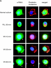
Relocalization of PAK4 to the cell membrane and colocalization of PAK4 and integrin αvβ5 in MCF-7 cells. (A) MCF-7 human breast carcinoma cells, under normal culture conditions or replated onto VN or PLL for indicated times, were stained for endogenous PAK4 using an anti-PAK4 pAb (green), for actin using phalloidin–rhodamine (red), and for nuclei by Hoechst (blue). Arrowheads indicate PAK4 localized to lamellipodia or ruffles after replating onto VN. (B) MCF-7 cells were replated onto VN and costained for endogenous PAK4 (green) and endogenous integrin αvβ5 (red). In the top panel (30 min after replating), integrin αvβ5 was found at lamellipodia (arrowheads). In the bottom panel (60 min after replating), integrin αvβ5 has started to form focal complexes at lamellipodia where it colocalized with PAK4 (arrowheads). Bars, 10 μm.
Figure 4.
Figure 4.
Relocalization of PAK4 to the cell membrane and colocalization of PAK4 and integrin αvβ5 in MCF-7 cells. (A) MCF-7 human breast carcinoma cells, under normal culture conditions or replated onto VN or PLL for indicated times, were stained for endogenous PAK4 using an anti-PAK4 pAb (green), for actin using phalloidin–rhodamine (red), and for nuclei by Hoechst (blue). Arrowheads indicate PAK4 localized to lamellipodia or ruffles after replating onto VN. (B) MCF-7 cells were replated onto VN and costained for endogenous PAK4 (green) and endogenous integrin αvβ5 (red). In the top panel (30 min after replating), integrin αvβ5 was found at lamellipodia (arrowheads). In the bottom panel (60 min after replating), integrin αvβ5 has started to form focal complexes at lamellipodia where it colocalized with PAK4 (arrowheads). Bars, 10 μm.
Figure 5.
Figure 5.
PAK4 translocation to lamellipodia by integrin ligation to VN does not depend on Cdc42 binding, integrin interaction, or PAK4 kinase activity. (A) Flag-tagged PAK4 mutants used for translocation studies. PAK4-L19, 22 lacks binding capacity to Cdc42/Rac. PAK4-M350 and PAK4-ΔIBD are both kinase dead, and PAK4-ΔIBD cannot bind integrin β5. (B) M21 cells were transfected with Flag-tagged wt PAK4, PAK4 mutants, or the control Flag-BAP vector. Cells were stained using an anti-Flag mAb (green), and stained for actin using phalloidin–rhodamine (red) and for nuclei using Hoechst (blue) before (B) or after (C) replating onto VN for 1 h. Arrows indicate the distribution in lamellipodia of PAK4 and PAK4 mutants. Bars, 20 μm. (D) Quantification of membrane-localized wt PAK4 or PAK4 mutants before and after replating onto VN for 1 h. Bars represent percent of cells with membrane-localized wt PAK4 or PAK4 mutants of the total cells counted and are expressed as mean ± SD. In a statistical evaluation comparing before and after cell replating onto VN, all PAK4 variants gave P < 0.05 (*) or P < 0.01 (**) by a paired t test. PAK4-L19, 22 localization to the membrane was also higher than wt PAK4 under normal culture conditions (P < 0.05 [*]).
Figure 5.
Figure 5.
PAK4 translocation to lamellipodia by integrin ligation to VN does not depend on Cdc42 binding, integrin interaction, or PAK4 kinase activity. (A) Flag-tagged PAK4 mutants used for translocation studies. PAK4-L19, 22 lacks binding capacity to Cdc42/Rac. PAK4-M350 and PAK4-ΔIBD are both kinase dead, and PAK4-ΔIBD cannot bind integrin β5. (B) M21 cells were transfected with Flag-tagged wt PAK4, PAK4 mutants, or the control Flag-BAP vector. Cells were stained using an anti-Flag mAb (green), and stained for actin using phalloidin–rhodamine (red) and for nuclei using Hoechst (blue) before (B) or after (C) replating onto VN for 1 h. Arrows indicate the distribution in lamellipodia of PAK4 and PAK4 mutants. Bars, 20 μm. (D) Quantification of membrane-localized wt PAK4 or PAK4 mutants before and after replating onto VN for 1 h. Bars represent percent of cells with membrane-localized wt PAK4 or PAK4 mutants of the total cells counted and are expressed as mean ± SD. In a statistical evaluation comparing before and after cell replating onto VN, all PAK4 variants gave P < 0.05 (*) or P < 0.01 (**) by a paired t test. PAK4-L19, 22 localization to the membrane was also higher than wt PAK4 under normal culture conditions (P < 0.05 [*]).
Figure 6.
Figure 6.
Dynamic localization of PAK4 in lamellipodia. MCF-7 human breast carcinoma cells stably transfected with EGFP–PAK4 or EGFP were plated onto VN and visualized by immunofluorescent microscopy after being allowed to attach for 30 min. Images were taken by a CCD camera every minute and the displayed cells are representative for the respective transfection. (A) EGFP–PAK4 is transiently localized in actively reshaping lamellipodia. (B) EGFP control exhibits only nuclear or perinuclear localization. Bars, 20 μm.
Figure 7.
Figure 7.
PAK4 stimulates integrin αvβ5–mediated cell migration. (A) MCF-7 cells transiently transfected with EGFP–PAK4 or EGFP control were analyzed for haptotactic cell migration toward VN in the presence or absence of normal mouse IgG and functional blocking mAbs LM609 (anti-αvβ3) or P1F6 (anti-αvβ5) (left panel) and toward collagen type I (right panel). Data represent the average of three independent experiments and were normalized to the transfection efficiency of individual vectors, as determined by flow cytometry. Statistical evaluation comparing EGFP to EGFP–PAK4 on VN gave P < 0.05 by t test. (B) Cell migration analyzed as in A of MCF-7 cells stably expressing EGFP–PAK4 or EGFP. All results are expressed as mean values ± SEM of three independent experiments using triplicate analysis in each experiment. Statistical evaluation by t test gave P < 0.05 for EGFP–PAK4 compared with EGFP on VN. (C) Overexpression of PAK4 decreases cell adhesion on VN. Cell attachment of MCF-7 cells stably expressing EGFP–PAK4 or EGFP at different coating concentrations of VN was determined. (D) Transient EGFP–PAK4 expression does not change the membrane expression levels of integrin αvβ5 in MCF-7 cells as measured by flow cytometry. αvβ5 expression was plotted versus EGFP content and the αvβ5 cell surface levels in EGFP-transfected cells (bottom left) was compared with that in EGFP–PAK4 cells (bottom right). Values indicate the mean fluorescence of αvβ5 staining in EGFP-positive cells. Unstained MCF-7 cells (top left) and staining without primary mAb (top right) were used to determine specificity and background.
Figure 7.
Figure 7.
PAK4 stimulates integrin αvβ5–mediated cell migration. (A) MCF-7 cells transiently transfected with EGFP–PAK4 or EGFP control were analyzed for haptotactic cell migration toward VN in the presence or absence of normal mouse IgG and functional blocking mAbs LM609 (anti-αvβ3) or P1F6 (anti-αvβ5) (left panel) and toward collagen type I (right panel). Data represent the average of three independent experiments and were normalized to the transfection efficiency of individual vectors, as determined by flow cytometry. Statistical evaluation comparing EGFP to EGFP–PAK4 on VN gave P < 0.05 by t test. (B) Cell migration analyzed as in A of MCF-7 cells stably expressing EGFP–PAK4 or EGFP. All results are expressed as mean values ± SEM of three independent experiments using triplicate analysis in each experiment. Statistical evaluation by t test gave P < 0.05 for EGFP–PAK4 compared with EGFP on VN. (C) Overexpression of PAK4 decreases cell adhesion on VN. Cell attachment of MCF-7 cells stably expressing EGFP–PAK4 or EGFP at different coating concentrations of VN was determined. (D) Transient EGFP–PAK4 expression does not change the membrane expression levels of integrin αvβ5 in MCF-7 cells as measured by flow cytometry. αvβ5 expression was plotted versus EGFP content and the αvβ5 cell surface levels in EGFP-transfected cells (bottom left) was compared with that in EGFP–PAK4 cells (bottom right). Values indicate the mean fluorescence of αvβ5 staining in EGFP-positive cells. Unstained MCF-7 cells (top left) and staining without primary mAb (top right) were used to determine specificity and background.

Similar articles

Cited by

References

    1. Abo, A., J. Qu, M.S. Cammarano, C. Dan, A. Fritsch, V. Baud, B. Belisle, and A. Minden. 1998. PAK4, a novel effector for Cdc42Hs, is implicated in the reorganization of the actin cytoskeleton and in the formation of filopodia. EMBO J. 17:6527–6540. - PMC - PubMed
    1. Bagrodia, S., and R.A. Cerione. 1999. PAK to the future. Trends Cell Biol. 9:350–355. - PubMed
    1. Bao, W., M. Thullberg, H. Zhang, A. Onischenko, and S. Strömblad. 2002. Cell attachment to the extracellular matrix induces proteasomal degradation of p21CIP1 via Cdc42/Rac1 signaling. Mol. Cell. Biol. 22:4587–4597. - PMC - PubMed
    1. Bar-Sagi, D., and A. Hall. 2000. Ras and Rho GTPases: a family reunion. Cell. 103:227–238. - PubMed
    1. Bodeau, A.L., A.L. Berrier, A.M. Mastrangelo, R. Martinez, and S.E. LaFlamme. 2001. A functional comparison of mutations in integrin β cytoplasmic domains: effects on the regulation of tyrosine phosphorylation, cell spreading, cell attachment, and β1 integrin conformation. J. Cell Sci. 114:2795–2807. - PubMed

Publication types

MeSH terms