. 2002 Sep 30;158(7):1299-309.
doi: 10.1083/jcb.200204056.

An extracellular site on tetraspanin CD151 determines alpha 3 and alpha 6 integrin-dependent cellular morphology

Affiliations

An extracellular site on tetraspanin CD151 determines alpha 3 and alpha 6 integrin-dependent cellular morphology

Alexander R Kazarov et al. J Cell Biol. .

Abstract

The alpha 3 beta 1 integrin shows strong, stoichiometric, direct lateral association with the tetraspanin CD151. As shown here, an extracellular CD151 site (QRD(194-196)) is required for strong (i.e., Triton X-100-resistant) alpha 3 beta 1 association and for maintenance of a key CD151 epitope (defined by monoclonal antibody TS151r) that is blocked upon alpha 3 integrin association. Strong CD151 association with integrin alpha 6 beta 1 also required the QRD(194-196) site and masked the TS151r epitope. For both alpha 3 and alpha 6 integrins, strong QRD/TS151r-dependent CD151 association occurred early in biosynthesis and involved alpha subunit precursor forms. In contrast, weaker associations of CD151 with itself, integrins, or other tetraspanins (Triton X-100-sensitive but Brij 96-resistant) were independent of the QRD/TS151r site, occurred late in biosynthesis, and involved mature integrin subunits. Presence of the CD151-QRD(194-196)-->INF mutant disrupted alpha 3 and alpha 6 integrin-dependent formation of a network of cellular cables by Cos7 or NIH3T3 cells on basement membrane Matrigel and markedly altered cell spreading. These results provide definitive evidence that strong lateral CD151-integrin association is functionally important, identify CD151 as a key player during alpha 3 and alpha 6 integrin-dependent matrix remodeling and cell spreading, and support a model of CD151 as a transmembrane linker between extracellular integrin domains and intracellular cytoskeleton/signaling molecules.


Figures

Figure 1.
Figure 1.
Schematic representations of wild-type and mutant CD151 and NAG-2. For all HA-tagged molecules, transmembrane (TM, hatched) and extracellular (EC) domains are indicated. Numbers in parentheses refer to last and first CD151 residues adjacent to downstream and upstream NAG-2 sequences, respectively. Asterisks indicate new mutants, distinct from those described previously (Yauch et al., 2000). Arrowheads mark positions of CD151 point mutations (C192Y and QRD194–196→INF) that completely abolish integrin association and the TS151r epitope. Anti-CD151 antibody staining (mAb 5C11, TS151r) of transfected Cos7 cells was assessed by flow cytometry. Neither mAb binds to monkey CD151 in Cos7 cells. Association with α3 integrin was detected in coimmunoprecipitation experiments using transiently transfected HT1080 cells. Key results were confirmed in Cos7 cells (e.g., Fig. 2). From 1% Triton X-100 cell lysates, CD151 mutants were immunoprecipitated using mAb anti–HA tag, and α3 integrin was immunoblotted using polyclonal antiserum D23.
Figure 2.
Figure 2.
CD151–INF194–196 loses integrin association. (A) Cos7 cells were transiently cotransfected with α3 integrin together with pZeoSV vector, wild-type CD151, or CD151–INF194–196. After 24– 36 h, cells were lysed in 1% Triton X-100 or 1% Brij 96 and immunoprecipitated with anti-CD151 5C11 mAb. Immune complexes were resolved in SDS-PAGE and immunoblotted with polyclonal anti-α3 antibodies or monoclonal anti-HA in reducing conditions. (B) Experiments were performed exactly as in A, except that Cos7 cells were cotransfected with α6 integrin and polyclonal anti-α6 antisera was used for immunoblotting in reducing conditions.
Figure 4.
Figure 4.
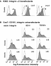
α3 and α6 integrins mask the TS151r epitope. (A) K562 cells were transiently transfected with α3 integrin or vector alone, and then the presence or absence of α3 was verified by flow cytometry using mAb IIF5. Cells lacking or expressing α3 were also stained using CD151 mAbs 5C11 (5) and TS151r (T) (middle and right, respectively). (B) Cos7 cells were transiently transfected with wild-type CD151, together with vector containing cDNA for α2 or α6 integrins, or vector alone. Cells were then stained for α2 integrin (mAb A2-IIE10), α6 subunit (mAb A6-ELE), or CD151 (mAbs 5C11 and TS151r). Negative control peaks were obtained using secondary antibody alone.
Figure 3.
Figure 3.
Loss of the TS151r epitope on CD151–INF194–196. Wild-type CD151, INF mutant, or pZeoSV vector alone were stably expressed in NIH3T3 cells, and then transfected cells were stained with mouse mAb 5C11 and TS151r and analyzed by flow cytometry as described (Zhang et al., 2002). Fluorescence histograms obtained using anti-CD151 mAb (5C11 and TS151r) are overlaid over negative control histograms obtained using cells transfected with vector alone (open histograms). Similar data was obtained to determine the presence or absence of TS151r and 5C11 binding to each tetraspanin structure shown in Fig. 1.
Figure 5.
Figure 5.
Association of CD151 with both α3 and α6 integrins during biosynthesis. 293 cells were transiently transfected with wild-type CD151-GFP alone or in combination with α3 or α6 integrins. Cells were pulsed with 0.5 mCi/ml [35S]methionine for 1 h and then chased in medium containing 5% dialyzed serum and 25× excess cold methionine for the indicated times (0, 1, 10, and 20 h) before lysis in RIPA buffer. Immunoprecipitation of CD151 was done using anti-GFP polyclonal antibody, and samples were resolved on 4–12% SDS-PAGE under nonreducing conditions and transferred to PVDF membrane. To detect 35S signal, membrane was exposed to BioMax MS film using BioMax Transcreen LE system. Coimmunoprecipitation of endogenous α3 or α6 was not observed (left lanes). In this regard, levels of transfected α3 were ∼5-fold greater than endogenous α3, and transfected α6 was present at ∼2-fold greater levels than endogenous α6 as estimated by flow cytometry (not depicted). Comparable levels of CD151-GFP were present for each transfectant as indicated by blotting with anti-GFP reagent (not depicted). Due to a large preexisting pool of intracellular β1, the integrin β1 subunit is not well labeled with 35S, particularly at early time points.
Figure 6.
Figure 6.
Cell lysis in Triton X-100 and Brij 96 yields distinct populations of CD151-associated integrins. HT1080 cells were labeled with [35S]methionine for 2.5, 5, 25, and 56 h. Cells were lysed in 1% Triton X-100 or Brij 96, and then immunoprecipitations of α3, CD151, and CD81 were performed using the indicated antibodies. Samples were resolved on 8–15% SDS-PAGE under reducing conditions and exposed to BioMax MS film. Note that during maturation, integrin α3 and α6 subunits are proteolytically cleaved, but fragments remain disulfide bonded. In reducing conditions, one can observe conversion of the ∼150-kD precursor form to fragments of ∼120 and 30 kD (not depicted). Labeled β1 subunit (110 kD), appearing at later time points, closely comigrates with 120 kD α subunit fragments. As seen elsewhere, CD81 associated bands of ∼120 kD from HT1080 cells (such as in panel e) have been clearly identified as corresponding to α3 and α6, with α3 being ∼2–3-fold more abundant than α6 in HT1080 cells (Berditchevski et al., 1996, 1997; Stipp et al., 2001).
Figure 7.
Figure 7.
QRD-independent CD151 associations. (A) Cos7 cells were transiently transfected with the indicated human CD151 constructs, lysed in the presence of 1% Triton X-100 or 1% Brij 96, and immunoprecipitated with anti-CD151 5C11 mAb. Resolved proteins were then immunoblotted using anti-CD151 mAb 1A5 in nonreducing conditions. Note that mAb 1A5 does not blot monkey CD151. (B) HT1080 cells were transiently transfected with the indicated HA-tagged CD151 constructs and immunoprecipitated using anti-HA mAb in a buffer containing 1% Brij 96. Immune complexes were resolved by SDS-PAGE and immunoblotted with anti-CD151 mAb 1A5 in nonreducing conditions, or anti-α3 polyclonal (D23), and anti-HA mAb in reducing conditions.
Figure 8.
Figure 8.
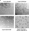
Functions of CD151–integrin complexes during cellular cable assembly. Human wild-type CD151 and CD151–INF194–196 mutant were highly expressed at comparable levels on the surface of Cos7 cells as determined by flow cytometry (not depicted). (a) Mock- or CD151-transfected cells (A–C) were plated on the surface of a thick layer of Matrigel in 5% FBS-DME at 5 × 10⁴ cells per well in a 24-well plate, analyzed using a ZEISS Axiovert 135 microscope, and photographed after 18 h as described previously (Zhang et al., 2002). In one case (D), anti-CD151 mAb 5C11 was added (at 7.5 μg/ml) at the beginning of the experiment. mAb 5C11 does not bind to endogenous monkey CD151. (b) Cos7 cell stable transfectants were treated as in part a, except that mAbs to integrin α6 (GoH3) or α3 (A3IIF5) were added (at 7.5 μg/ml) at the beginning of the experiment. The GoH3 and A3IIF5 mAbs do recognize monkey α6 and α3 integrins. Note that compared with α3 levels in mock-transfected Cos7 cells (MFI = 222), α3 levels in mutant or wild-type CD151-transfected cells varied by ≤1.2-fold. Compared with α6 levels in mock-transfected Cos7 cells (MFI = 55), α6 levels in mutant or wild-type CD151-transfected cells varied by ≤1.9-fold. Previously we learned that twofold differences in α6 levels had essentially no effect on static cell adhesion or on cell cable formation on Matrigel (Zhang et al., 2002). (c) Stable NIH3T3 cell transfectants were grown in 5% FBS-DME at 105 cells per well (24 well plate) on the surface of Matrigel for 20 h before photographs were taken.
Figure 8.
Figure 8.
Functions of CD151–integrin complexes during cellular cable assembly. Human wild-type CD151 and CD151–INF194–196 mutant were highly expressed at comparable levels on the surface of Cos7 cells as determined by flow cytometry (not depicted). (a) Mock- or CD151-transfected cells (A–C) were plated on the surface of a thick layer of Matrigel in 5% FBS-DME at 5 × 10⁴ cells per well in a 24-well plate, analyzed using a ZEISS Axiovert 135 microscope, and photographed after 18 h as described previously (Zhang et al., 2002). In one case (D), anti-CD151 mAb 5C11 was added (at 7.5 μg/ml) at the beginning of the experiment. mAb 5C11 does not bind to endogenous monkey CD151. (b) Cos7 cell stable transfectants were treated as in part a, except that mAbs to integrin α6 (GoH3) or α3 (A3IIF5) were added (at 7.5 μg/ml) at the beginning of the experiment. The GoH3 and A3IIF5 mAbs do recognize monkey α6 and α3 integrins. Note that compared with α3 levels in mock-transfected Cos7 cells (MFI = 222), α3 levels in mutant or wild-type CD151-transfected cells varied by ≤1.2-fold. Compared with α6 levels in mock-transfected Cos7 cells (MFI = 55), α6 levels in mutant or wild-type CD151-transfected cells varied by ≤1.9-fold. Previously we learned that twofold differences in α6 levels had essentially no effect on static cell adhesion or on cell cable formation on Matrigel (Zhang et al., 2002). (c) Stable NIH3T3 cell transfectants were grown in 5% FBS-DME at 105 cells per well (24 well plate) on the surface of Matrigel for 20 h before photographs were taken.
Figure 9.
Figure 9.
Morphology of CD151 transfectants on Matrigel. Cells were seeded onto coverslips with a thin coating of Matrigel (1/30 dilution of stock solution, according to manufacturer's instruction) and incubated in serum-free DME at 37°C. Photographs of spread cells were taken using a 20× lens of ZEISS Axiovert 135 microscope. Photos of individual cells are representative of the majority of CD151 wild-type (CD151wt) and CD151–INF transfectants at each of the time points.
Figure 10.
Figure 10.
Quantitation of cell spreading on Matrigel and fibronectin. Spread cells were defined as cells that lost phase bright appearance and increased their surface contact area by at least twofold. Coverslips were coated with Matrigel (as in the legend to Fig. 9) or with fibronectin (10 μg/ml). For each curve, ≥50 or more cells were counted. At 50 min and beyond, spreading on Matrigel was identical for both cell types (at ∼95%; not depicted). Results shown are representative of three separate experiments.

Similar articles

Cited by

References

    1. Anton, E.S., J.A. Kreidberg, and P. Rakic. 1999. Distinct functions of alpha3 and alpha(v) integrin receptors in neuronal migration and laminar organization of the cerebral cortex. Neuron. 22:277–289. - PubMed
    1. Belkin, A.M., and M.A. Stepp. 2000. Integrins as receptors for laminins. Microsc. Res. Tech. 51:280–301. - PubMed
    1. Bell, E., B. Ivarsson, and C. Merrill. 1979. Production of a tissue-like structure by contraction of collagen lattices by human fibroblasts of different proliferative potential in vitro. Proc. Natl. Acad. Sci. USA. 76:1274–1278. - PMC - PubMed
    1. Berditchevski, F. 2001. Complexes of tetraspanins with integrins: more than meets the eye. J. Cell Sci. 114:4143–4151. - PubMed
    1. Berditchevski, F., G. Bazzoni, and M.E. Hemler. 1995. Specific association of CD63 with the VLA-3 and VLA-6 integrins. J. Biol. Chem. 270:17784–17790. - PubMed

Publication types