. 2002 Oct 1;544(Pt 1):211-23.
doi: 10.1113/jphysiol.2002.022368.

Activation of different vestibular subnuclei evokes differential respiratory and pressor responses in the rat

Affiliations

Activation of different vestibular subnuclei evokes differential respiratory and pressor responses in the rat

Fadi Xu et al. J Physiol. .

Abstract

Activation of the vestibular system can either increase or decrease ventilation. The objectives of the present study were to clarify whether these different responses are the result of activating different vestibular subnuclei, by addressing three questions. Do neurones within the medial, lateral and spinal vestibular nuclei (VN(M), VN(L) and VN(S), respectively) function differently in respiratory modulation? Is the ventral medullary nucleus gigantocellularis (NGC) required to fully express the VN-mediated respiratory responses? Is glutamate, by acting on N-methyl-D-aspartic acid (NMDA) receptors in the vestibular subnuclei, capable of modulating respiration? In anaesthetized, tracheotomized and spontaneously breathing rats, electrical stimuli (< 10 s) applied in the VN(L) and VN(S) significantly elevated ventilation by 35 % and 30 % (P < 0.05), respectively. However, VN(M) stimulation produced statistically significant (P < 0.05) changes that differed depending upon the stimulation site: either ventilatory inhibition (by 40 % in 57 % of the trials) or excitation (by 55 % in 43 % of trials), and which were often accompanied by a pressor response. These electrical-stimulation-evoked cardiorespiratory responses were almost eliminated following microinjection of ibotenic acid into the stimulation sites (P < 0.05) or bilaterally into the NGC (P < 0.05). As compared to vehicle, microinjection of NMDA into the unilateral VN(M), VN(L) and VN(S) significantly increased ventilation to 74 %, 58 % and 60 % (P < 0.05), respectively, with no effect on arterial blood pressure. These data suggest that neurones within the vestibular subnuclei play different roles in cardiorespiratory modulation, and that the integrity of the NGC is essential for the full expression of these VN-mediated responses. The evoked respiratory excitatory responses are probably mediated by glutamate acting on NMDA receptors, whereas the neurotransmitters involved in VN(M)-mediated respiratory inhibition and hypertension remain unknown.


Figures

Figure 1
Figure 1. Experimental recordings of ventilatory and arterial blood pressure (ABP) responses to stimulation of the medial (VNM), lateral (VNL) and spinal (VNS) vestibular nuclei
Experimental recordings of ventilatory and ABP responses to stimulation of the VNM (A and B), VNS (C) and VNL (D). In each panel, the traces from the top to bottom are ABP, tidal volume (VT), airflow and tracheal pressure (Ptr). Note that the thin and thick bars indicate stimulation at 100 and 150 Hz, respectively.
Figure 2
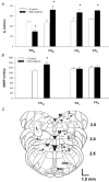
Figure 2. Group data showing the effects of stimulation of the vestibular subnuclei on cardiorespiratory responses
The responses of minute ventilation (VI) and mean arterial blood pressure (MABP) are illustrated in A and B, respectively. The number of animals used for VNM, VNL and VNS stimulation was 27, 11 and 10, respectively; data are presented as the mean ±s.e.m.; *P < 0.05 between the data obtained before and during electrical stimulation. C, placements of the stimulating electrode are mapped schematically in a group of cross-sections of the brainstem 2.5-3.5 mm rostral to the obex according to the rat brain atlas (Paxinos & Watson, 1986). M, L and S represent the VNM, VNL and VNS, respectively; Amb, nucleus ambiguus; NGC, gigantocellular nucleus. The locations of the electrodes referenced to stereotaxic coordinates and those derived from Chicago Sky Blue staining are represented by open and filled symbols, respectively. The triangles and squares denote the inhibitory and excitatory respiratory responses, respectively (complete overlap occurred at six sites: two inhibitory and four excitatory).
Figure 3
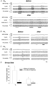
Figure 3. Effects of ibotenic acid (IA) microinjection into the vestibular subnuclei on the cardiorespiratory responses evoked by local electrical stimulation of VNM (A), VNL (B) and VNS (C)
In each panel, the traces from the top to bottom are ABP, VT, airflow and Ptr. The thin and thick bars indicate stimulation at 100 and 200 Hz, respectively. Group data comparing cardiorespiratory responses before and after local IA injection are depicted in D. The number of rats used for stimulation of the VNM, VNL and VNS was six, two and two, respectively; the data are presented as means ±s.e.m.; *P < 0.05 between the responses to vestibular nucleus (VN) stimulation and control (without electrical stimulation); †P < 0.05 between the responses before and after IA injection. Please note that the MABP data were collected from six rats that exhibited a pressor response to VNM stimulation at 150 Hz.
Figure 4
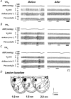
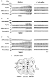
Figure 4. Comparison of the evoked cardiorespiratory responses immediately before and 2 h after NGC lesions
A-C, the cardiorespiratory responses to stimulation of the VNM, VNS and VNL. In each panel, the traces from the top to bottom are ABP, VT, airflow and Ptr. Note that the first, second and third (if presented) stimulation denoted by bars indicate a stimulus intensity of 100, 150 and 200 Hz, respectively. Representative areas stained by Chicago Sky Blue (IA) injections are illustrated schematically (one side) in D, where the measurements given at the bottom are the distances rostral to the obex. LPGi, lateral paragigantocellular nucleus (see Paxinos & Watson, 1986).
Figure 5
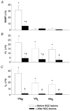
Figure 5. Group data showing the effects of NGC lesions on VN-stimulation-induced changes in MABP (A), VT (B) and respiratory frequency (fR; C)
The numbers of rats for stimulation of the VNM, VNL and VNS were 20, 8 and 7, respectively; the data are presented as means ±s.e.m.; *P < 0.05 compared to control, †P < 0.05 between the responses before and after bilateral IA injection into the NGC.
Figure 6
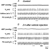
Figure 6. Cardiorespiratory responses to stimulation of the VNM before and 2 h after vehicle (100 nl) injection into the bilateral NGC in a rat
In each panel, the traces from the top to bottom are ABP, VT, airflow and Ptr. The thin and thick bars indicate stimulation at 100 and 200 Hz, respectively.
Figure 7
Figure 7. The cardiorespiratory responses to microinjection of N-methyl-d-aspartic acid (NMDA) into the VNM (A), VNL (B) and VNS (C)
The immediate responses and their recovery (2 min after NMDA injection) are illustrated in the left and right columns, respectively. In each panel, the traces from the top to bottom are ABP, VT, airflow and Ptr. The horizontal bars represent the duration of microinjection of 50 nl NMDA into the given subnucleus. Injection sites stained by Chicago Sky Blue are marked in D, in which the numbers listed are the distances rostral to the obex. py, pyramidal tract (see Paxinos & Watson, 1986).
Figure 8
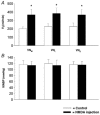
Figure 8. Group data presenting the effects of NMDA injection in the vestibular subnuclei on ventilation (A) and ABP (B)
The number of animals in which NMDA was injected into the VNM, VNL and VNS was nine, nine and eight, respectively; data are presented as means ±s.e.m.; *P < 0.05 compared to control.

Similar articles

Cited by

References

    1. Bassal M, Bianchi AL. Inspiratory onset or termination induced by electrical stimulation of the brain. Respiration Physiology. 1982;50:23–40. - PubMed
    1. Billig I, Hartge K, Card JP, Yates BJ. Transneuronal tracing of neural pathways controlling abdominal musculature in the ferret. Brain Research. 2001;912:24–32. - PubMed
    1. Bystrzycka EK, Nail BS. The source of the respiratory drive to nasolabialis motoneurons in the rabbit: a HRP study. Brain Research. 1983;266:183–191. - PubMed
    1. Chan SH. Arterial pressure- and cardiac rhythm-related single-neuron activities in the nucleus reticularis gigantocellularis (NRGC) Journal of the Autonomic Nervous System. 1983;13:99–109. - PubMed
    1. Chen LW, Yung KK, Chan YS. Co-localization of NMDA receptors and AMPA receptors in neurons of the vestibular nuclei of rats. Brain Research. 2000;884:87–97. - PubMed

Publication types

MeSH terms

LinkOut - more resources