. 2002 Oct 1;544(Pt 1):225-36.
doi: 10.1113/jphysiol.2002.024505.

Length-dependent activation in three striated muscle types of the rat

Affiliations

Length-dependent activation in three striated muscle types of the rat

John P Konhilas et al. J Physiol. .

Abstract

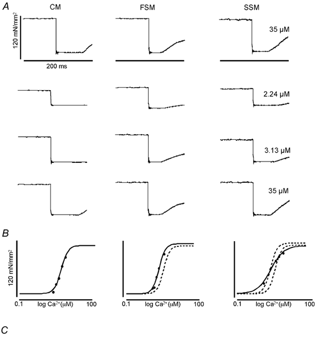
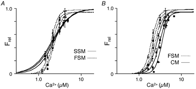
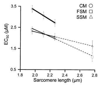
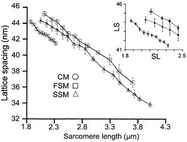
The process whereby sarcomere length modulates the sensitivity of the myofilaments to Ca(2+) is termed length-dependent activation. Length-dependent activation is a property of all striated muscles, yet the relative extent of length-dependent activation between skeletal muscle and cardiac muscle is unclear. Although length-dependent activation may be greater in fast skeletal muscle (FSM) than in slow skeletal muscle (SSM), there has not been a well controlled comparison of length-dependent activation between skeletal muscle and cardiac muscle (CM). Accordingly, we measured sarcomere length-dependent properties in skinned soleus (SSM), psoas (FSM) and ventricular trabeculae (CM) of the rat under carefully controlled conditions. The free Ca(2+)-force relationship was determined at sarcomere lengths (SL) of 1.95 microm, 2.10 microm and 2.25 microm and fitted to a modified Hill equation. FSM and SSM were more sensitive to Ca(2+) than CM. Length-dependent activation was ordered as CM > FSM > SSM. Cooperativity as measured by the Hill coefficient of the Ca(2+)-force relationship was not significantly different between CM and FSM, both of which exhibited greater cooperativity than SSM. SL did not significantly alter this parameter in each muscle type. To establish whether the observed differences can be explained by alterations in interfilament spacing, we measured myofilament lattice spacing (LS) by synchrotron X-ray diffraction in relaxed, skinned muscle preparations. LS was inversely proportional to SL for each muscle type. The slope of the SL-LS relationship, however, was not significantly different between striated muscle types. We conclude that (1) length-dependent activation differs among the three types of striated muscle and (2) these differences in the length-dependent properties among the striated muscle types may not solely be explained by the differences in the response of interfilament spacing to changes in muscle length in relaxed, skinned isolated muscle preparations.


Figures

Figure 1
Figure 1. Method used to measure active force development under maximal and submaximal activation in rat cardiac trabeculae (CM), rat psoas (FSM), and soleus (SSM) muscle fibres
A, original recordings of force, normalized as force (unit area−1, at the indicated [Ca2+] (in μM) to the right of the tracings for each striated muscle type (SL = 2.10 μm). A quick release was used to determine zero force level (see Methods). The quick release step size depended on muscle type and [Ca2+]; it was 10–20 % of muscle length. Calibrations as indicated. B, Ca2+-force relationship, constructed from data obtained as illustrated in A. The continuous lines indicate the Hill fit to the data obtained from the indicated muscle type (above each data column); the dotted lines represent the Hill fit obtained from CM (middle panel), or CM and FSM (right panel). C, typical CCD images of X-ray diffraction patterns obtained from each muscle type at the same SL (SL = 2.10 μm) indicative of the change in lateral separation of the myofilaments in each muscle. The 1,0 and 1,1 equatorial reflections are as indicated.
Figure 2
Figure 2. Ca2+-dependent force development in skinned cardiac trabeculae, psoas and soleus muscle fibres
A, Ca2+-force relationship at three sarcomere lengths (1.95 μm (circles), 2.10 μm (squares), 2.25 μm (triangles)) of psoas (FSM; open symbols) and soleus (SSM; filled symbols) single muscle fibres. B, Ca2+-force relationship at the same three sarcomere lengths as in A of psoas (FSM; open symbols) and cardiac trabeculae (CM; filled symbols). The data were normalized to Ca2+-saturated (maximum) force at each SL.
Figure 3
Figure 3. Relationship between Ca2+ sensitivity (EC50) and sarcomere length in skinned cardiac (CM), psoas (FSM), and soleus (SSM) muscle fibres
The relationship in each muscle type was linear over the range of sarcomere lengths studied (1.95 μm, 2.10 μm and 2.25 μm) representing the working range of the heart. The slope of these relationships reflects the magnitude of length-dependent activation; the steeper the slope, the greater the shift in Ca2+ sensitivity with changes in length. The dashed line represents the linear extrapolation of these data to SL = 2.75 μm consistent with the Ca2+ sensitivity (EC50) obtained in a separate group skeletal muscle fibres at that length.
Figure 4
Figure 4. Averaged lattice spacing as a function of sarcomere length
Shown are the sarcomere length-lattice spacing relationships determined from the three striated muscle types studied. These relationships were obtained by placing the sarcomere length on 0.05 μm (CM) or 0.10 μm (FSM and SSM) intervals into bins and averaging the average value per fibre bin. Inset, sarcomere length-lattice spacing relationships scaled to the working range of cardiac muscle.

References

    1. Akella AB, Ding XL, Cheng R, Gulati J. Diminished Ca2+ sensitivity of skinned cardiac muscle contractility coincident with troponin T-band shifts in the diabetic rat. Circulation Research. 1995;76:600–606. - PubMed
    1. Allen DG, Kentish JC. The cellular basis of the length-tension relation in cardiac muscle. Journal of Molecular and Cellular Cardiology. 1985;17:821–840. - PubMed
    1. Allen DG, Kurihara S. The effects of muscle length on intracellular calcium transients in mammalian cardiac muscle. Journal of Physiology. 1982;327:79–94. - PMC - PubMed
    1. Allen JD, Moss RL. Factors influencing the ascending limb of the sarcomere length-tension relationship in rabbit skinned muscle fibres. Journal of Physiology. 1987;390:119–136. - PMC - PubMed
    1. Arteaga GM, Palmiter KA, Leiden JM, Solaro RJ. Attenuation of length dependence of calcium activation in myofilaments of transgenic mouse hearts expressing slow skeletal troponin I. Journal of Physiology. 2000;526:541–549. - PMC - PubMed

Publication types

LinkOut - more resources