. 2002 Oct 29;99(22):14548-53.
doi: 10.1073/pnas.212319799. Epub 2002 Sep 30.

Selenomethionine regulation of p53 by a ref1-dependent redox mechanism

Affiliations

Selenomethionine regulation of p53 by a ref1-dependent redox mechanism

Young R Seo et al. Proc Natl Acad Sci U S A. .

Abstract

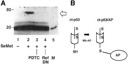
The cancer chemopreventive properties of selenium compounds are well documented, yet little is known of the mechanism(s) by which these agents inhibit carcinogenesis. We show that selenium in the form of selenomethionine (SeMet) can activate the p53 tumor suppressor protein by a redox mechanism that requires the redox factor Ref1. Assays to measure direct reduction/oxidation of p53 showed a SeMet-dependent response that was blocked by a dominant-negative Ref1. By using a peptide containing only p53 cysteine residues 275 and 277, we demonstrate the importance of these residues in the SeMet-induced response. SeMet induced sequence-specific DNA binding and transactivation by p53. Finally, cellular responses to SeMet were determined in mouse embryo fibroblasts wild-type or null for p53 genes. The evidence suggests that the DNA repair branch of the p53 pathway was activated. The central relevance of DNA repair to cancer prevention is discussed.


Figures

Fig 1.
Fig 1.
Reduction response of p53 protein to SeMet. (A) Reduction/oxidation of full-length p53. H1299 cells (p53-null) were transiently transfected with a wild-type p53 expression plasmid. Cells were treated with 20 μM SeMet and p53 protein reduction/oxidation assayed (19). Briefly, oxidized cysteine sulfhydryl residues were detected by lysis in the presence of NEM, which reacts with and blocks free sulfhydryls. Disulfide linkages then were reduced by DTT and labeled with the biotinylation reagent MPB; p53 was immunoprecipitated with Ab421, and biotinylated p53-detected by blotting with streptavidin HRP. Alternatively, p53 sulfhydryls were labeled directly with MPB to detect reduced p53 forms. The lanes reflect SeMet and other treatments. Lane 1, untreated cells; lane 2, cells treated with SeMet exhibit a p53 redox response; lane 3, abrogation of the p53 reduction response by dominant–negative Ref1 mutant (Ref-DN); lane 4, abrogation of the p53 reduction response by PDTC, known to oxidize p53 cysteines (positive control). Reduced and oxidized p53 cysteine residues were inversely correlated in SeMet-treated cells. Immune complexes were probed with Ab421 to ascertain equivalent gel loading. (B) Diagram of redox chemistry methodology used in A. Sulfhydryl groups were first covalently blocked by reaction with NEM (S-H to S-R modification), then disulfide-reduced by DTT and labeled with MPB for subsequent streptavidin detection (19). Alternatively, p53 sulfhydryls were labeled directly with MPB to detect reduced p53 forms.
Fig 2.
Fig 2.
Redox regulation of p53 Cys-275 and/or Cys-277. (A) H1299 cells (p53-null) were transiently transfected with an expression plasmid encoding the carboxyl-terminal one-third of p53 protein. The carboxyl-terminal p53 peptide (ct-p53) migrates as a 20-kDa band. Reaction with maleimide-activated alkaline phosphatase (MA-AP) causes ct-p53 to migrate as a 70-kDa ct-p53/alkaline phosphatase (AP) conjugate, detected by Ab421 to p53. The lanes reflect SeMet and other treatments. Lane 1, untreated cells; lane 2, cells treated with SeMet exhibit a ct-p53 reduction response; lane 3, abrogation of the reduction response by PDTC; lane 4, abrogation of the reduction response by dominant–negative Ref1 mutant (Ref-DN); lane 5, peptide alone without modification by MA-AP, used to mark the position of unmodified peptide (M). (B) Diagram of redox chemistry used in A. A gel-shift assay was used to detect directly reduced forms of p53, specifically, of p53 residues 275 and/or 277. Free sulfhydryl groups were labeled with MA-AP, which forms a covalent linkage of sulfhydryl groups to AP. The resulting ctp53/AP conjugate migrates at approximately 70 kDa.
Fig 3.
Fig 3.
Promotion of p53 1620+ conformation in response to SeMet. H1299 cells transiently transfected with wild-type p53 were treated with 20 μM SeMet for 15 h, and immune complexes were collected by using Ab1620, a conformation-sensitive epitope present on active p53. The lanes reflect SeMet and other treatments. Lane 1, untreated cells; lane 2, cells treated with SeMet exhibit 1620+ p53; lane 3, abrogation of the 1620+ response by Ref-DN mutant; lane 4, abrogation of the 1620+ response by PDTC. (A) Immunoblots of p53 protein in 1620+ immune complexes. Total p53 was detected by blotting of lysates with antibody D01 without immunoprecipitation. (B) Quantification of three independent experiments, mean ± SD. P < 0.02, by Wilcoxon rank-sum test.
Fig 4.
Fig 4.
Transcriptional activation of p53 by SeMet. (A) EMSA showing binding of p53 to a p53-responsive gene sequence. Extracts were prepared from H1299 cells transiently transfected with p53 expression plasmids. Lane 1, untreated cells; lane 2, cells treated with 20 μM SeMet for 15 h showed enhanced sequence-specific binding of p53; lane 3, cells treated with PDTC abrogates p53 activation by SeMet; lane 4, cells treated with UV radiation (positive control); lane 5, absence of binding to mutant oligonucleotide sequence (negative control; Santa Cruz Biotechnology). Arrow denotes the position of the Ab421-p53 supershifted complex. (B) Dependence of supershifted complex formation on Ab421. Lane 6, absence of Ab421; lane 7, addition of Ab421; lane 8, addition of Ab421 and DO1. Open arrow denotes the position of the doubly shifted complex. (C) Activation of a p53-responsive reporter gene by SeMet. H1299 cells were cotransfected with p53 expression vectors and pG13-CAT, a reporter construct that carries 13 p53 binding sites to measure p53 activation. Lane 1, untreated cells; lane 2, treatment with SeMet enhances p53-dependent transcription; lane 3, abrogation by PDTC; lane 4, abrogation by Ref-DN mutant; lane 5, lack of pG13-CAT activation by a p53-mutant (Val-143 → Ala; negative control). Quantification was by a CAT enzyme ELISA assay. Relative CAT enzyme units are shown; mean ± SD from five independent experiments (P < 0.01 by t test, using SIGMAPLOT software).
Fig 5.
Fig 5.
Cellular responses to SeMet involving p53. (A) SeMet protected p53 wild-type but not p53-null cells from UV radiation. MEF cells were treated with 20 μM SeMet for 15 h, then with UV radiation at the indicated doses. Cell yield was determined after 7 days. SeMet protected wild-type MEFs, whereas p53-null cells remained UV sensitive. SeMet had no effect on survival of either cell line in the absence of DNA damage. Shown is the mean ± SD of three independent determinations (P < 0.02 by Wilcoxon rank-sum test). (B) DNA repair induction by SeMet in p53 wild-type but not p53 mutant cells. An isogenic matched pair of MCF7 cell lines were used (ref. 17). DNA repair was measured by HCR of a UV-damaged reporter gene. Cells carrying a p53 mutant (Val-143 → Ala) did not show induction of the HCR response (17); PDTC abrogated SeMet induction of the HCR response. Shown is the mean ± SD of five independent experiments (P < 0.01 by t test using SIGMAPLOT software).

Comment in

  • New careers for antioxidants.
    Brash DE, Havre PA. Brash DE, et al. Proc Natl Acad Sci U S A. 2002 Oct 29;99(22):13969-71. doi: 10.1073/pnas.232574399. Epub 2002 Oct 21. Proc Natl Acad Sci U S A. 2002. PMID: 12391310 Free PMC article. Review. No abstract available.

Similar articles

Cited by

References

    1. Ip C. (1981) Cancer Res. 41, 4386-4390. - PubMed
    1. Reddy B. S., Sugie, S., Maryyama, H., el-Bayoumy, K. & Marra, P. (1987) Cancer Res. 47, 5901-5904. - PubMed
    1. Nelson M. A., Porterfield, B. W., Jacobs, E. T. & Clark, L. C. (1999) Semin. Urol. Oncol. 17, 91-96. - PubMed
    1. Lu J., Jiang, C., Kaeck, M., Ganther, H., Vadhanavikit, S., Ip, C. & Thompson, H. J. (1995) Biochem. Pharmacol. 50, 213-219. - PubMed
    1. Sinha R., Said, T. K. & Medina, D. (1996) Cancer Lett. (Shannon, Irel.) 107, 277-284. - PubMed

Publication types