

Crystal structure of human calcineurin complexed with cyclosporin A and human cyclophilin
- PMID: 12357034
- PMCID: PMC129706
- DOI: 10.1073/pnas.212504399
Crystal structure of human calcineurin complexed with cyclosporin A and human cyclophilin
Abstract
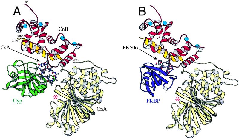
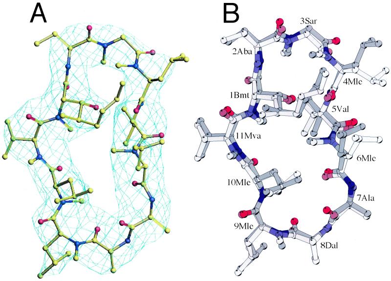
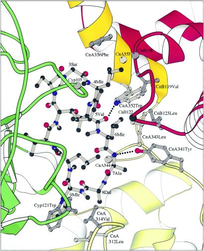
Calcineurin (Cn), a Ca(2+)/calmodulin-dependent Ser/Thr protein phosphatase, is an important participant in signaling pathways that activate T cells. It is the target of the immunosuppressive drugs cyclosporin A (CsA) and FK506. These drugs bind proteins known as cyclophilin (Cyp) and FK506-binding protein, respectively, and the drug-protein complexes in turn inhibit Cn. We report the crystal structure of a Cyp/CsA/Cn ternary complex, determined to a resolution of 3.1 A. Residues 3-9 of CsA, particularly N-methyl leucines 4 and 6, and Trp-121 of Cyp form a composite surface for interaction with Cn. The hydrophobic interface buries two hydrogen bonds. The structure accounts clearly for the effects of mutations in Cn on CsA-resistance and for the way modifications of CsA alter immunosuppressive activity.
Figures
Similar articles
-
Crystal structure of calcineurin-cyclophilin-cyclosporin shows common but distinct recognition of immunophilin-drug complexes.Proc Natl Acad Sci U S A. 2002 Sep 17;99(19):12037-42. doi: 10.1073/pnas.192206699. Epub 2002 Sep 6. Proc Natl Acad Sci U S A. 2002. PMID: 12218175 Free PMC article.
-
A proposed molecular model for the interaction of calcineurin with the cyclosporin A-cyclophilin A complex.Bioorg Med Chem. 1999 Jul;7(7):1389-402. doi: 10.1016/s0968-0896(99)00072-3. Bioorg Med Chem. 1999. PMID: 10465413
-
Structures of calcineurin and its complexes with immunophilins-immunosuppressants.Biochem Biophys Res Commun. 2003 Nov 28;311(4):1095-102. doi: 10.1016/s0006-291x(03)01537-7. Biochem Biophys Res Commun. 2003. PMID: 14623295 Review.
-
Three-dimensional structure and actions of immunosuppressants and their immunophilins.FASEB J. 1995 Jan;9(1):63-72. doi: 10.1096/fasebj.9.1.7529736. FASEB J. 1995. PMID: 7529736 Review.
-
Substitution in position 3 of cyclosporin A abolishes the cyclophilin-mediated gain-of-function mechanism but not immunosuppression.J Biol Chem. 2004 Jan 23;279(4):2470-9. doi: 10.1074/jbc.M304754200. Epub 2003 Oct 28. J Biol Chem. 2004. PMID: 14583619
Cited by
-
Optimizing PK properties of cyclic peptides: the effect of side chain substitutions on permeability and clearance().Medchemcomm. 2012 Oct;3(10):1282-1289. doi: 10.1039/C2MD20203D. Medchemcomm. 2012. PMID: 23133740 Free PMC article.
-
Expanding the PP2A Interactome by Defining a B56-Specific SLiM.Structure. 2016 Dec 6;24(12):2174-2181. doi: 10.1016/j.str.2016.09.010. Epub 2016 Oct 27. Structure. 2016. PMID: 27998540 Free PMC article.
-
SPLINTS: small-molecule protein ligand interface stabilizers.Curr Opin Struct Biol. 2016 Apr;37:115-22. doi: 10.1016/j.sbi.2016.01.004. Epub 2016 Jan 30. Curr Opin Struct Biol. 2016. PMID: 26829757 Free PMC article. Review.
-
Peptidylprolyl Isomerases as In Vivo Carriers for Drugs That Target Various Intracellular Entities.Biomolecules. 2017 Sep 29;7(4):72. doi: 10.3390/biom7040072. Biomolecules. 2017. PMID: 28961224 Free PMC article. Review.
-
Calcineurin is required for Candida albicans to survive calcium stress in serum.Infect Immun. 2005 Sep;73(9):5767-74. doi: 10.1128/IAI.73.9.5767-5774.2005. Infect Immun. 2005. PMID: 16113294 Free PMC article.
References
-
- Aramburu J., Rao, A. & Klee, C. B. (2000) Curr. Top. Cell. Regul. 36, 237-295. - PubMed
-
- Griffith J. P., Kim, J. L., Kim, E. E., Sintchak, M. D., Thomson, J. A., Fitzgibbon, M. J., Fleming, M. A., Caron, P. R., Hsiao, K. & Navia, M. A. (1995) Cell 82, 507-522. - PubMed
-
- Kissinger C. R., Parge, H. E., Knighton, D. R., Lewis, C. T., Pelletier, L. A., Tempczyk, A., Kalish, V. J., Tucker, K. D., Showalter, R. E., Moomaw, E. W., et al. (1995) Nature 378, 641-644. - PubMed
-
- Fliri H., Baumann, G., Enz, A., Kallen, J., Luyten, M., Mikol, V., Movva, R., Quesniaux, V., Schreier, M., Walkinshaw, M., et al. (1993) Ann. N.Y. Acad. Sci. 696, 47-53. - PubMed
-
- Mikol V., Kallen, J., Pflugl, G. & Walkinshaw, M. D. (1993) J. Mol. Biol. 234, 1119-1130. - PubMed
Publication types
MeSH terms
Substances
Associated data
- Actions
LinkOut - more resources
Full Text Sources
Other Literature Sources
Molecular Biology Databases
Miscellaneous
