

doi: 10.1159/000149474.
Isolation and morphology of the internal component of human coronavirus, strain 229E
- PMID: 1235860
- PMCID: PMC7179545
- DOI: 10.1159/000149474
Item in Clipboard
Isolation and morphology of the internal component of human coronavirus, strain 229E
Intervirology.
1975.
Abstract
Biochemical studies on human coronavirus, strain 229E, indicate that the RNA is present in the virion in association with protein as a ribonucleoprotein (RNP) complex. Morphological studies have revealed that this nucleocapsid is probably a continuous helical RNP.
Figures
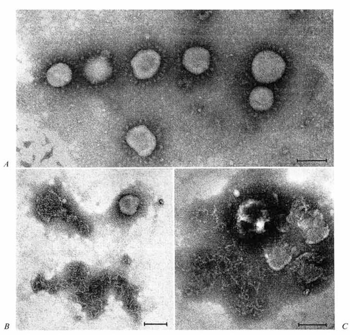
A Freshly prepared virus. Morphology is typical of the Coronaviridae. B Particles in three stages of disruption, seen after 24-hour storage at 4; upper right particle without petal-like projections is typical of many; upper left shows particle apparently losing internal component, but no structural detail is evident: bottom center shows typical discrete tangle of threadlike strands, 8-9 nm in diameter. C Preparation as in B, showing particle releasing filamentous material. Bars represent 100 nm in all micrographs.
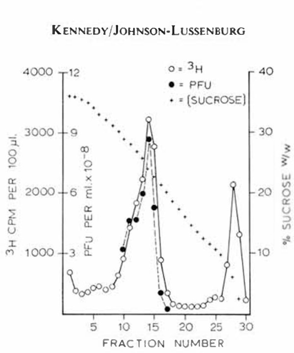
Distribution of 3H-label and infectivity in sucrose gradient after rate-zonal centrifugation of HCV/229E.
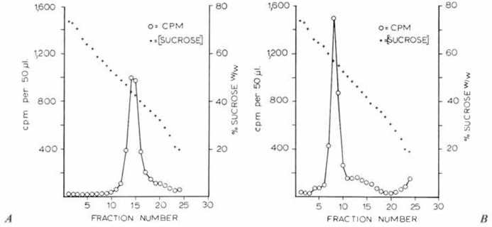
Centrifugation of (A) control and (B) NP-40-trcated 3H-uridine-labelled HCV/229E in 25-75% w/w sucrose gradients at 170,000 g for 22 h.
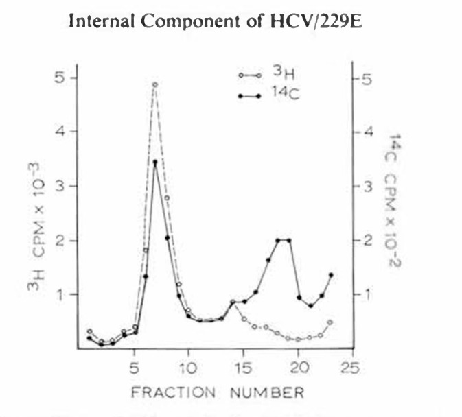
Distribution of 3H and 14C activity in 25-75% sucrose gradient centrifuged at 170,000 g for 22 h. HCV/229E was labelled with 3H-uridine and 14C-amino acids and treated with NP-40 before centrifugation.
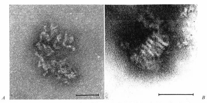
A Typical appearance of particles seen in 3H-labelled peak of gradient fractions of NP-40-trcated HCV/229E preparations. B Tightly coiled particle, also from NP-40-trcated HCV/229E samples. Bar represents 100 nm.
References
-
- Apostolov K., Flewett T.H. Internal structure of influenza virus. Virology. 1965;26:506–508. - PubMed
-
- Horse R.W., Waterson A.P. A helical structure in mumps, Newcastle disease and Sendai viruses. J. molec. Biol. 1960;2:75–77.
-
- Howatson A.F., Whitmore G.F. The development and structure of vesicular stomatitis virus. Virology. 1962;16:466–478. - PubMed
-
- Simpson R.W., Hauser R.E. Structural components of vesicular stomatitis virus. Virology. 1966;29:654–667. - PubMed
-
- Nowinski R.C., Lloyd J.O., Sarkar N.H., Moore D.H. Common properties of the oncogenic RNA viruses (oncornaviruses) Virology. 1970;42:1152–1157. - PubMed
MeSH terms
Substances
LinkOut - more resources
Full Text Sources
