. 2002 Oct 2:3:18.
doi: 10.1186/1471-2156-3-18. Epub 2002 Oct 2.

The human L-threonine 3-dehydrogenase gene is an expressed pseudogene

Affiliations

The human L-threonine 3-dehydrogenase gene is an expressed pseudogene

Alasdair J Edgar. BMC Genet. .

Abstract

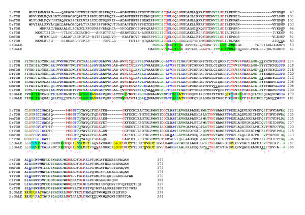
Background: L-threonine is an indispensable amino acid. One of the major L-threonine degradation pathways is the conversion of L-threonine via 2-amino-3-ketobutyrate to glycine. L-threonine dehydrogenase (EC 1.1.1.103) is the first enzyme in the pathway and catalyses the reaction: L-threonine + NAD+ = 2-amino-3-ketobutyrate + NADH. The murine and porcine L-threonine dehydrogenase genes (TDH) have been identified previously, but the human gene has not been identified.

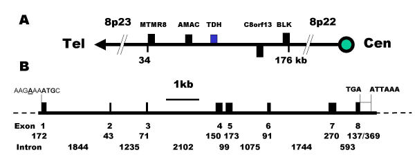
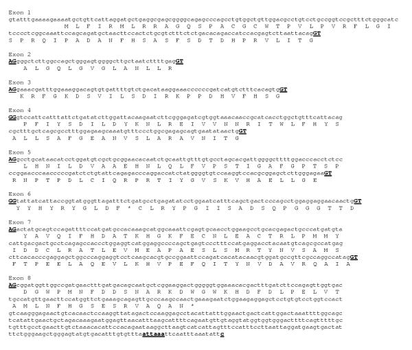
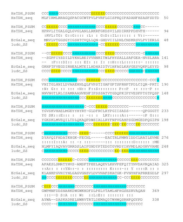
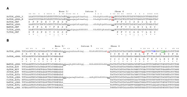
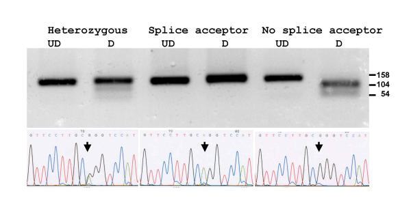
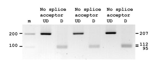
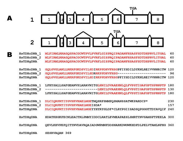
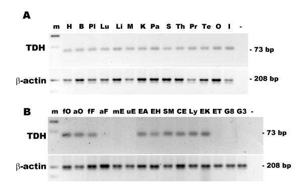
Results: The human TDH gene is located at 8p23-22 and has 8 exons spanning 10 kb that would have been expected to encode a 369 residue ORF. However, 2 cDNA TDH transcripts encode truncated proteins of 157 and 230 residues. These truncated proteins are the result of 3 mutations within the gene. There is a SNP, A to G, present in the genomic DNA sequence of some individuals which results in the loss of the acceptor splice site preceding exon 4. The acceptor splice site preceding exon 6 was lost in all 23 individuals genotyped and there is an in-frame stop codon in exon 6 (CGA to TGA) resulting in arginine-214 being replaced by a stop codon. These truncated proteins would be non-functional since they have lost part of the NAD+ binding motif and the COOH terminal domain that is thought to be involved in binding L-threonine. TDH mRNA was present in all tissues examined.

Conclusions: The human L-threonine 3-dehydrogenase gene is an expressed pseudogene having lost the splice acceptor site preceding exon 6 and codon arginine-214 (CGA) is mutated to a stop codon (TGA).


Figures

Figure 1
Figure 1
Chromosomal localisation and gene structure of the human TDH gene. (A) The human TDH gene is located at 8p23 and the neighbouring genes are: myotubularin related protein 8, MTMR8: acyl-malonyl condensing enzyme, AMAC: threonine dehydrogenase, TDH: hypothetical protein, C8orf13; B lymphoid tyrosine kinase, BLK. Genes encoded by + and - stands are shown above and below the line respectively. The telomeric and centromeric directions are indicated. (B) The human TDH gene spans 10 kb and consists of 8 exons. The putative initiation methionine codon, stop codon and poly-adenylation signal are shown. Closed boxes indicate the ORF and the sizes (in bp) of the exons and introns are indicated.
Figure 2
Figure 2
Sequence and translation of the human TDH exons. A comparison of the human genomic sequence with the murine TDH gene enabled the identification of 8 homologous exons on clone RP11-110L10. A translation of the exons is shown below the nucleotide sequence. The acceptor and donor splice sites (AG &GT) at the ends of each exon are shown in uppercase and underlined. The potential polyadenylation signal and site (attaaa and c) are shown in bold and underlined.
Figure 3
Figure 3
Clustal alignment of the predicted human protein with other eukaryotic L-threonine dehydrogenase (TDH) protein sequences and 2 UDP-galactose 4-epimerase sequences (GALE). The species and the derivation of the sequences are as follows: Homo sapiens, HsTDH, genomic DNA (accession No. AC011959); HsGALE, cDNA (NP_000394); Sus scrofa, (pig), Ss, cDNA (AY095535); Mus musculus, mouse, Mm, cDNA (AY116662); Danio rerio (zebrafish), Dr, ESTs (electronic contigous sequence); Takifugu rubripes (puffer fish), Tr, genomic DNA, (AF411956); Ciona intestinalis (a tunicate), Ci, ESTs; Drosophila melanogaster, Dm, genomic DNA gene CG5955 and ESTs; Caenorhabditis elegans, Ce, genomic DNA (U64847) and ESTs; Escherichia coli, EcTDH (P07913) and EcGALE (AAC73846). The locations of the exon/exon boundaries are shown on the translated protein as underlined residues. Conserved residues are indicated by a (*), strongly similar residues by a (:) and weakly similar residues by a (.). Residues that are common to at least two protein sequences are shown in bold. The in-frame stop codon in the human sequence is indicated by a red (#). Gale protein residues in contact with the ligand nicotinamide-adenine-dinucleotide are highlighted in green and those residues in contact with the sugar ligands, uridine-5'-diphosphate-mannose and uridine-5'-diphosphate-4-deoxy-4-fluoro-alpha-d-galactose are highlighted in yellow. Those residues that have been shown to contact both NAD and sugar are highlighted in light blue [26-29].
Figure 4
Figure 4
Predicted secondary structure of human "ancestral" TDH protein was determined using the Psi-Pred program and was aligned with that of the crystal structure of uridine diphosphogalactose-4-epimerase protein (GALE) from Escherichia coli[29,31] using 3D-PSSM. The labels are: human "ancestral" TDH protein predicted secondary structure, HsTDH_PSSM; human "ancestral" TDH protein sequence, HsTDH_seq; E. coli GALE protein sequence, EcGale_seq; E. coli GALE protein secondary structure, 1udc_SS; alpha-helix, H, highlighted in light blue; beta-sheet, E, highlighted in yellow; c = turn, coil or loop. Identical residues are shown and additionally, a ":" indicates positive equivalence.
Figure 5
Figure 5
Comparison of the human TDH gene sequences in the region of exon 4 and 6 acceptor sites with those of other species. (A) The splice donor and acceptor sites are indicated (gt and ag respectively). There is a splice site mutation in exon 4 in one of the human genomic sequences (gg, coloured red). The translations of the genes are shown below the DNA sequences (ORF). The species and accession numbers are: Homo sapiens, HsTDH_gDNA_A (AF131216); HsTDH_gDNA_B (AC011959); Mus musculus, MmTDHgDNA (14.54000001-55000000 supercontig, Sanger Centre, UK) and puffer fish, Takifugu rubripes, TrTDH_gDNA (AF411956). (B) There is an in-frame stop codon (TGA, coloured blue) and a splice site mutation in exon 6 in the human genomic sequence (AF131216). A cryptic splice site (ag, coloured red) was utilised in cDNA clone 1 that altered the reading frame, resulting in another stop codon (TGA, coloured green). The species and accession numbers are: pig, Sus scrofa, SsTDH_cDNA (AY095535); MmTDH_cDNA (AY116662); rat, Rattus norvegicus, RmTDH_EST (BF557448); chicken, Gallus gallus, GgTDH_EST (BM491791); western clawed frog, Silurana tropicalis, StTDH_EST (AL638064); zebrafish, Danio rerio, DrTDH_ESTs; the tunicate, Ciona intestinalis, CiTDH_ESTs; fruit fly, Drosophila melanogaster, DmTDH_gDNA (AAF51607); the nematode, Caenorhabditis elegans, CeTDH_ESTs and the euglenoid, Trypanosoma brucei, TbTDH_gDNA (AC084047). There are no introns in this region of the fly, nematode and trypanosome genes (__). Identical nucleotides and residues are shown by *, strongly similar residues by: and weakly similar residues by (.).
Figure 6
Figure 6
Amplicons from individuals with different genotypes for the exon 4 splice acceptor site SNP were digested with the restriction enzyme AciI (upper panel) and DNA sequenced (lower panel). Undigested samples, UD and digested samples, D. Arrows indicate the polymorphic nucleotide. In an individual homozygous for the splice acceptor site (AG, middle panel) no AciI digest site is present. In an individual homozygous for loss of the splice acceptor site (GG, right panel) the 158 bp amplicon is cut into fragments of 104 and 54 bp. In an individual heterozygous for the AG splice acceptor site (RG, left panel) both alleles are present and the amplicon is only partially digested.
Figure 7
Figure 7
Amplicons from 3 individuals for the exon 6 splice acceptor site were digested with the restriction enzyme Hsp92II. All amplicons were digested showing loss of the splice site. Undigested samples, UD and digested samples, D.
Figure 8
Figure 8
The human TDH pseudogene is expressed. (A) Clones from 2 individuals were sequenced and mapped to the TDH gene. Clone 1 skipped exon 6, and the resulting frame-shift generated a premature stop codon in exon 7 (TGA). Clone 2 skipped exon 4 and utilised a cryptic splice site in exon 6, and the resulting frame-shift generated a premature stop codon in exon 6. (B) The translation of clone 1 (exon 6 skipped) encoded a truncated 230 residue ORF. The skipped exon 4 in clone 2 does not alter the reading frame. However, the use of a cryptic splice site in exon 6, results in a premature stop codon in exon 6. Stop codons are indicated by *.
Figure 9
Figure 9
Expression of the TDH pseudogene in human tissues and cell types. (A) Expression of the TDH pseudogene in human tissues. The lanes are: 100 bp marker, m; heart, H; brain, B; placenta, Pl; lung, Lu; liver, L; skeletal muscle, M; kidney, K; pancreas, Pa; spleen, S; thymus; Th; prostate, Pr; testis, Te; ovary, O; small intestine, I; negative control, (-). (B) Expression of the TDH pseudogene in human cells. RT-PCR for TDH (top panel) and β-actin (bottom panel). The cell types examined were: foetal osteoblast, fO; adult osteoblasts, aO; pulmonary foetal fibroblast, HFL-1, fF; adult fibroblasts, aF; placental microvascular endothelial, mE; umbilical vein endothelial, uE; alveolar epithelial adenocarcinoma, A549, EA; bronchial epithelial adenocarcinoma, H322, EH; bronchial smooth muscle, SM; colorectal adenocarcinoma, CaCo2, CE; Epstein-Barr-transformed lymphocyte Ly; erythroleukaemia, K562, EK; erythroleukaemia TF1, ET; glioma, U87, G8; glioma, U373, G3; and negative control (-).

References

    1. Strain AJ, Neuberger JM. A bioartificial liver – state of the art. Science. 2002;295:1005–1009. doi: 10.1126/science.1068660. - DOI - PubMed
    1. Tsiaoussis J, Newsome PN, Nelson LJ, Hayes PC, Plevris JN. Which hepatocyte will it be? Hepatocyte choice for bioartificial liver support systems. Liver Transpl. 2001;7:2–10. doi: 10.1053/jlts.2001.20845. - DOI - PubMed
    1. Enard W, Khaitovich P, Klose J, Zollner S, Heissig F, Giavalisco P, Nieselt-Struwe K, Muchmore E, Varki A, Ravid R, et al. Intra- and interspecific variation in primate gene expression patterns. Science. 2002;296:340–343. doi: 10.1126/science.1068996. - DOI - PubMed
    1. Cross DM, Bayliss MK. A commentary on the use of hepatocytes in drug metabolism studies during drug discovery and development. Drug Metab Rev. 2000;32:219–240. doi: 10.1081/DMR-100100574. - DOI - PubMed
    1. Dabos KJ, Nelson LJ, Bradnock TJ, Parkinson JA, Sadler IH, Hayes PC, Plevris JN. The simulated microgravity environment maintains key metabolic functions and promotes aggregation of primary porcine hepatocytes. Biochim Biophys Acta. 2001;1526:119–130. doi: 10.1016/S0304-4165(01)00097-6. - DOI - PubMed

MeSH terms