. 2002 Oct 15;99(21):13465-70.
doi: 10.1073/pnas.202287499. Epub 2002 Oct 2.

Protonation of non-Watson-Crick base pairs and encapsidation of turnip yellow mosaic virus RNA

Affiliations

Protonation of non-Watson-Crick base pairs and encapsidation of turnip yellow mosaic virus RNA

Hugo H J Bink et al. Proc Natl Acad Sci U S A. .

Abstract

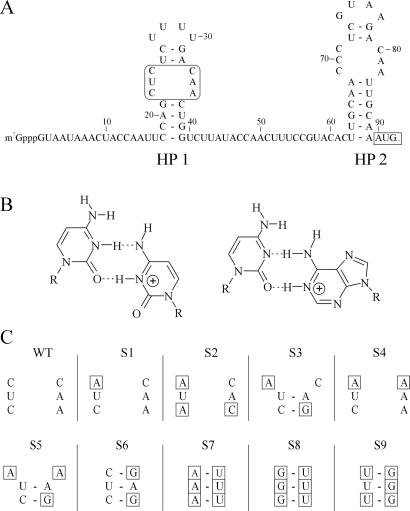
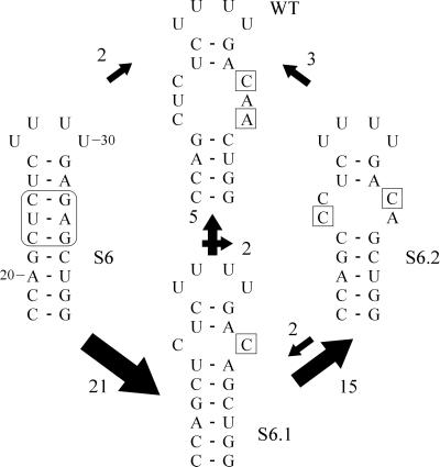
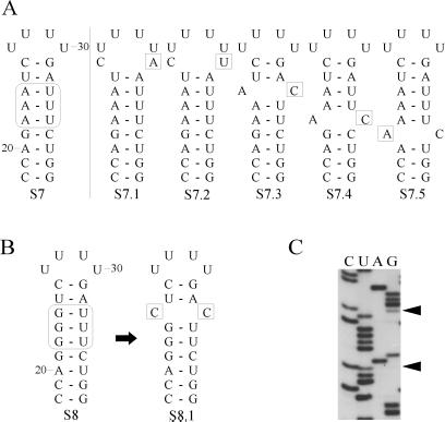
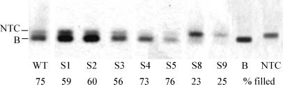
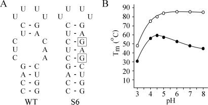
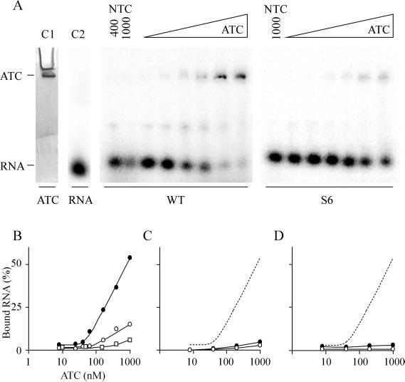
The 5' UTR of turnip yellow mosaic virus RNA contains two conserved hairpins with internal loops consisting of C.C and C.A mismatches. In this article, evidence is presented indicating that the 5' proximal hairpin functions as an encapsidation initiation signal. Extensive mutagenesis studies on this hairpin and sequencing of virus progeny showed a clear preference for C.C and C.A mismatches within the internal loop. The importance of these mismatches lies in their pH-dependent protonation and stable base pair formation. Encapsidation efficiency was found to be severely affected for several mutants lacking the protonatable mismatches in the internal loop of the 5' proximal hairpin. Furthermore, gel mobility-shift assays were performed with various RNA hairpins and empty capsids with a hole. Protonatable hairpins containing C.C and/or C.A pairs were found to bind specifically to the interior of the protein shell under acidic conditions (pH 4.5) in the presence of spermidine. Based on these results we propose that this binding of protonated cytosines to the coat protein of turnip yellow mosaic virus may represent a new motif in RNA-protein interactions.


Figures

Figure 1
Figure 1
Overview of the substitution mutations made in the internal loop of the 5′ proximal hairpin of TYMV RNA. (A) The 5′ UTR of TYMV RNA including the two hairpins containing the protonatable internal loops (HP1 and HP2). The mutagenesis site and the start codon of movement protein (MP) are boxed. (B) Proposed structure of a protonated C⋅C and C⋅A mismatch. Note the identical positioning of the two exocyclic amino groups in the deep groove. (C) Overview of the substitution mutants S1–S9 used in this study. Base substitutions in the internal loop, as compared with WT, are boxed.
Figure 2
Figure 2
Reversion pathway for mutant S6. Changes in the 5′ UTR sequence were monitored over four rounds of infection. Differences in sequence as compared with the original mutant are boxed. The arrows and their size indicate the direction and frequency of accumulation of changes in the internal loop region. After the first round of infection 21 of 25 cases first generated S6.1, having one C⋅C mismatch at the same position as the WT. In the successive rounds of infection the majority of revertants S6.1 (75%) evolved to form S6.2, including a C⋅C as well as a C⋅A mismatch. Numbering of the sequence is according to the nucleotide position in the viral RNA genome.
Figure 3
Figure 3
Observed reversion for mutant S7 (A) and S8 (B). Differences in sequence as compared with the original mutation are boxed. (C) Autoradiogram showing that the progeny of mutant S8 contains a mixed sequence at the positions indicated by arrowheads.
Figure 4
Figure 4
Determination of the ratio of filled and empty particles of WT and various mutant virus preparations by means of agarose gel electrophoresis. Lane B represents purified B component. The values indicate the average percentage of filled particles for every mutant.
Figure 5
Figure 5
UV melting as a function of pH. (A) RNA fragments used. The boxes indicate substitutions in mutant S6 as compared with WT. (B) Influence of pH on midpoint temperature Tm. ●, WT 5′ proximal hairpin; ○, mutant S6 hairpin.
Figure 6
Figure 6
Gel mobility-shift assay. (A) Binding experiments using WT HP1 and mutant hairpin S6 at different concentrations of ATC, ranging from 8 to 1,000 nM. Binding conditions include 50 mM Na acetate, pH 4.5, and 3 mM spermidine. The control lane (C1) containing ATC only was stained separately with Coomassie brilliant blue. In lane C2 32P-labeled WT HP1 was loaded. The control lanes NTC contain either 1,000 and 400 nM, or 1,000 nM only of NTC in the binding reactions with HP1 and hairpin S6, respectively. (B) Plot of the relative amounts of bound RNA as shown in A for the WT HP1 (●) and mutant S6 (○). Hairpin Y (□), consisting of Watson–Crick base pairs but with a different sequence, was used as a control. (C) Binding conditions: 50 mM Na acetate, pH 4.5. The dashed line is the WT HP1 shift as found in B. (D) Binding conditions: 50 mM Na cacodylate, pH 7.0, and 3 mM spermidine.

Similar articles

Cited by

References

    1. Canady M A, Larson S B, Day J, McPherson A. Nat Struct Biol. 1996;3:771–781. - PubMed
    1. Giegé R. Proc Natl Acad Sci USA. 1996;93:12078–12081. - PMC - PubMed
    1. Hellendoorn K, Michiels P J A, Buitenhuis R, Pleij C W A. Nucleic Acids Res. 1996;24:4910–4917. - PMC - PubMed
    1. Hellendoorn K. Ph.D. thesis. Leiden, The Netherlands: Leiden University; 1997.
    1. Hellendoorn K, Verlaan P W G, Pleij C W A. J Virol. 1997;71:8774–8779. - PMC - PubMed

Publication types

LinkOut - more resources