. 2002 Oct 15;99(21):13753-8.
doi: 10.1073/pnas.212221199. Epub 2002 Oct 2.

Functional evidence for the mediation of diabetogenic T cell responses by HLA-A2.1 MHC class I molecules through transgenic expression in NOD mice

Affiliations

Functional evidence for the mediation of diabetogenic T cell responses by HLA-A2.1 MHC class I molecules through transgenic expression in NOD mice

Michele P Marron et al. Proc Natl Acad Sci U S A. .

Abstract

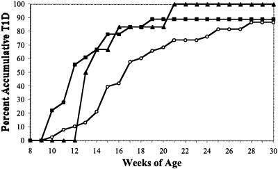
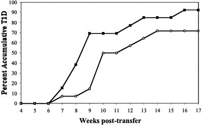
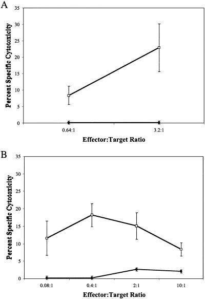
Particular major histocompatibility complex (MHC) class II alleles clearly contribute to T cell-mediated autoimmune type 1 diabetes (T1D) in both humans and nonobese diabetic (NOD) mice. However, studies in NOD mice indicate MHC class I-restricted T cell responses are also essential to T1D development. In humans, epidemiological studies have suggested that some common class I alleles, including HLA-A2.1 (A*02011), may confer increased susceptibility to T1D when expressed in conjunction with certain class II alleles. We show here that when HLA-A2.1 molecules are transgenically expressed in NOD mice, A2-restricted T cell responses arise against pancreatic beta cells, leading to an earlier onset of T1D. The accelerated onset of T1D in the NOD.HLA-A2.1 transgenic mice is not due to nonspecific effects of expressing a third class I molecule, because a stock of NOD mice transgenically expressing HLA-B27 class I molecules showed no such acceleration of T1D, but rather were significantly protected from disease. These findings provide the first functional evidence that certain human MHC class I molecules can contribute to the development of T1D.


Figures

Figure 1
Figure 1
Accelerated onset of diabetes in HLA-A2.1 transgenic NOD mice. Female NOD (open circles, n = 38) and NOD.HLA-A2.1 mice homozygous (filled squares, n = 18) or heterozygous (filled triangles, n = 6) for the transgene were monitored 30 weeks for development of diabetes. T1D development was significantly increased in NOD mice transgenically expressing HLA-A2.1 molecules in homozygous (P = 0.02) or heterozygous (P = 0.04) state.
Figure 2
Figure 2
HLA-A2.1 expression restores T1D susceptibility to NOD.β2mnull.hβ2m mice. The HLA-A2.1 transgene was crossed onto the NOD.β2mnull.hβ2m stock. Female NOD mice (open circles, n = 38) and NOD.β2mnull.hβ2m mice expressing (filled squares, n = 18) or not expressing (open triangles, n = 11) HLA-A2.1 were monitored 30 weeks for development of diabetes. T1D development in NOD.β2mnull.hβ2m mice is significantly decreased compared with NOD controls (P = 0.002). Expression of the HLA-A2.1 transgene in NOD.β2mnull.hβ2m.HLA-A2.1 mice reconstitutes T1D development to an incidence not significantly different from standard NOD mice (P = 0.4).
Figure 3
Figure 3
Accelerated adoptive transfer of T1D by NOD.HLA-A2.1 splenocytes. Splenocytes (1 × 10⁷) from 7- to 8-week-old female donors were transferred by i.v. injection into the indicated scid recipients. NOD.HLA-A2.1 splenocytes were injected into either NOD-scid.HLA-A2.1 (open triangles, n = 20) or NOD-scid (filled squares, n = 24) female recipients. NOD-scid recipients were also repopulated with standard NOD female splenocytes (open circles, n = 18). Recipient mice were monitored 17 weeks after repopulation for development of diabetes. T1D was transferred to NOD-scid recipients more rapidly by splenocytes from NOD.HLA-A2.1 than standard NOD donors (P = 0.03).
Figure 4
Figure 4
HLA-A2.1 expression does not increase the positive selection of diabetogenic T cells by using murine MHC class I molecules for their effector function. NOD bone marrow cells (5 × 10⁶) were injected into lethally irradiated NOD or NOD.HLA-A2.1 recipients. Six weeks after marrow reconstitution, splenocytes from the NOD and NOD.HLA-A2.1 chimeras were found to contain equal proportions of both CD4 T cells (32.4% vs. 30.6%, respectively) and CD8 T cells (11.3% vs. 10.5%, respectively). At this time, 1 × 10⁷ pooled splenocytes from each chimera group were transferred by i.v. injection into NOD-scid recipients. NOD-scid recipients were monitored 17 weeks after repopulation for development of diabetes. TID developed more slowly (P = 0.05) in recipients of NOD T cells that had matured in the presence (open circles, n = 14) rather than the absence (filled squares, n = 13) of HLA-A2.1 molecules on thymic epithelium.
Figure 5
Figure 5
Lysis of NOD-scid or NOD-scid.HLA-A2.1 β cell targets by isolated autoreactive CD8 T lymphocytes. Lymphocytes cultured from islets of two 10-week-old (A) or one 6-week-old (B) NOD.HLA-A2.1 female mouse were used as effectors in a 51Cr release assay at the indicated effector:target ratios. β cell targets were isolated from lymphocyte-deficient NOD-scid (filled circles) or NOD-scid.HLA-A2.1 (open squares) mice.

References

    1. Serreze D V, Leiter E H. Curr Dir Autoimmun. 2001;4:31–67. - PubMed
    1. Todd J A, Wicker L S. Immunity. 2001;15:387–395. - PubMed
    1. Sheehy M J. Diabetes. 1992;41:123–129. - PubMed
    1. Todd J A. Springer Semin Immunopathol. 1992;14:33–58. - PubMed
    1. Prochazka M, Serreze D V, Worthen S M, Leiter E H. Diabetes. 1989;38:1446–1455. - PubMed

Publication types

LinkOut - more resources