. 2002 Oct 15;99(21):13583-8.
doi: 10.1073/pnas.212483099. Epub 2002 Oct 2.

Higher order arrangement of the eukaryotic nuclear bodies

Affiliations

Higher order arrangement of the eukaryotic nuclear bodies

I-Fan Wang et al. Proc Natl Acad Sci U S A. .

Abstract

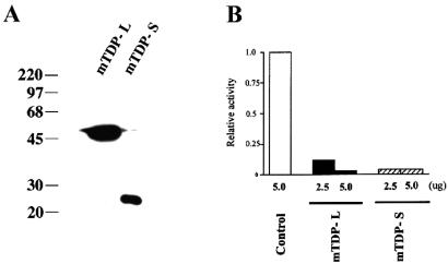
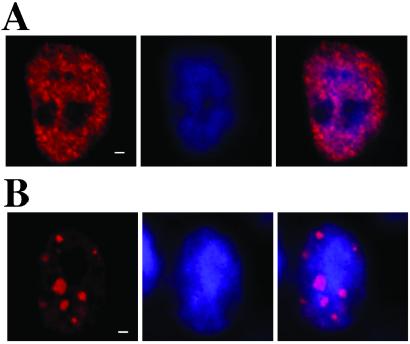
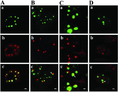
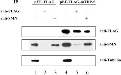
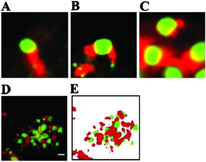
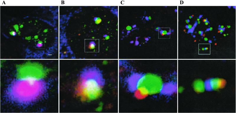
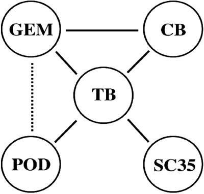
The nuclei of eukaryotic cells consist of discrete substructures. These substructures include the nuclear bodies, which have been implicated in a number of biological processes such as transcription and splicing. However, for most nuclear bodies, the details of involvement in these processes in relation to their three-dimensional distributions in the nucleus are still unclear. Through the analysis of TDP, a protein functional in both transcriptional repression and alternative splicing, we have identified a new category of nuclear bodies within which the TDP molecules reside. Remarkably, the TDP bodies (TBs) colocalize or overlap with several different types of nuclear bodies previously suggested to function in transcription or splicing. Of these nuclear bodies, the Gemini of coiled bodies (GEM) seems to associate with TB through the interaction between survival motor neuron (SMN) protein and TDP. Furthermore, TB sometimes appears to be the bridge of two or more of these other nuclear bodies. Our data suggest the existence of a hierarchy and possibly functional arrangement of the nuclear bodies within the eukaryotic nuclei.


Figures

Figure 1
Figure 1
Sequence alignment of hTDP-43, mTDP-L, and mTDP-S. The nucleotide and amino acid sequences of the cDNAs encoding hTDP-43, mTDP-L, and mTDP-S, respectively, are aligned for comparison. The mTDP-L sequence was put together through sequencing of the RT-PCR products and of a mouse EST clone (GenBank accession no. AA116656). The two RRM domains conserved in all three proteins are underlined, and the RNP-1 and RNP-2 motifs within the two domains are boxed. The glycine-rich region present in hTDP-43 and mTDP-L, but not in mTDP-S, are indicated with italic letters. The polyadenylation signal near the 3′ ends of the hTDP-L and mTDP-L cDNAs are indicated with large, underlined letters.
Figure 2
Figure 2
Transcriptional repression by mTDP-L and mTDP-S. 293 cells were cotransfected with 1 μg of the reporter plasmids (pG0-α-Luc) and increasing amounts (2.5 and 5 μg) of the expression plasmids (pEF-FLAG-mTDP-L or pEF-FLAG-mTDP-S) or the vector. At 24 h posttransfection, the cells were harvested for analysis of expression of the FLAG-tagged mTDP-L and mTDP-S by Western blotting (A) and for an assay of their effects on the expression of the cotransfected reporter plasmids (B).
Figure 3
Figure 3
Cellular immunostaining patterns of FLAG-mTDP-L and FLAG-mTDP-S. 293 cells were transfected with pEF-FLAG-mTDP-L (A) or pEF-FLAG-mTDP-S (B) and analyzed by immunostaining with anti-FLAG antibody (red, A and B Left) or with the Hoechst DNA dye (blue, A and B Center). The merged images are shown in A and B Right. (Scale bar, 1 μm.)
Figure 4
Figure 4
Immunostaining patterns of eGFP-mTDP-S-expressing cells. 293 cells cotransfected with pCMV-eGFP-mTDP-S and pEF-FLAG-mTDP-S (A) or transfected with pCMV-eGFP-mTDP-S alone (BD) were immunostained with antibodies against the FLAG epitope (A), coilin (B), SMN (C), or PML (D). The images of the green fluorescence from eGFP-mTDP-S (AaDa) and the red fluorescence from the secondary antibodies (AbDb) are merged in AcDc. The overlapping/colocalized regions exhibit the yellow color, as shown in Ac and exemplified with the arrows in BcDc. (Scale bar, 1 μm.)
Figure 5
Figure 5
Cross-IP of SMN and TDP. Extracts from 293 cells transfected with pEF-FLAG vector (lanes 1–3) or pEF-FLAG-mTDP-S (lanes 4–6) were analyzed by Western blotting using anti-FLAG, anti-SMN, or antitubulin after IP with anti-FLAG (lanes 2 and 5) or anti-SMN (lane 3 and 6). Extract without the IP step was used as the control in lanes 1 and 4. The detections of the SMN band in lane 5 by anti-SMN and of the FLAG-mTDP-S band in lane 6 by anti-FLAG indicate the interaction between SMN and FLAG-mTDP-S in vivo.
Figure 6
Figure 6
Interconnection of TB and SC35 speckles. 293 cells transfected with pCMV-eGFP-mTDP-S were analyzed by immunostaining with anti-SC35. Examples of merged images of TB (green) and SC35 (red) are shown in AD. The image in D is from one section of a nucleus with highly reticulated interconnection of TB and SC35 speckles. The 3D reconstruction of this nucleus is shown in E. (Scale bar in D, 2 μm.)
Figure 7
Figure 7
Interconnection among three different nuclear bodies. 293 cells transfected with pCMV-eGFP-mTDP-S were double-stained with different pairs of antibodies: (A) anti-coilin (red) and anti-SMN (blue); (B) anti-SMN (blue) and anti-PML (red); (C) anti-coilin (red) and anti-SC35 (blue); and (D) anti-PML (red) and anti-SC35 (blue). Note that the merged color is yellow for green plus red, purple for red plus blue, sky blue for green plus blue, and white for green plus red plus blue. The boxed regions in AD Upper are magnified in AD Lower. (Scale bar in A Upper, 1 μm.)
Figure 8
Figure 8
Association of TB with other nuclear bodies. The associations among five different nuclear bodies (TB, GEM, CB, SC35, and POD) are presented schematically. The association between GEM and CB has been described also in ref. . The less frequently observed association between POD and GEM is indicated by the dotted line.

Similar articles

Cited by

References

    1. Lamond A I, Earnshaw W C. Science. 1998;280:547–553. - PubMed
    1. Misteli T, Spector D L. Curr Opin Cell Biol. 1998;10:323–331. - PubMed
    1. Matera A G. Trends Cell Biol. 1999;9:302–309. - PubMed
    1. Misteli T. Science. 2001;291:843–847. - PubMed
    1. Cremer T, Cremer C. Nat Rev. 2001;2:292–301. - PubMed

Publication types

MeSH terms

LinkOut - more resources