

The function of bursts of spikes during visual fixation in the awake primate lateral geniculate nucleus and primary visual cortex
- PMID: 12361982
- PMCID: PMC129798
- DOI: 10.1073/pnas.212500599
The function of bursts of spikes during visual fixation in the awake primate lateral geniculate nucleus and primary visual cortex
Abstract
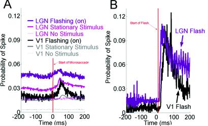
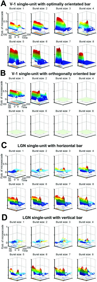
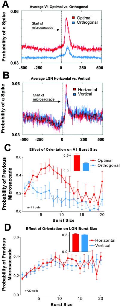
When images are stabilized on the retina, visual perception fades. During voluntary visual fixation, however, constantly occurring small eye movements, including microsaccades, prevent this fading. We previously showed that microsaccades generated bursty firing in the primary visual cortex (area V-1) in the presence of stationary stimuli. Here we examine the neural activity generated by microsaccades in the lateral geniculate nucleus (LGN), and in the area V-1 of the awake monkey, for various functionally relevant stimulus parameters. During visual fixation, microsaccades drove LGN neurons by moving their receptive fields across a stationary stimulus, offering a likely explanation of how microsaccades block fading during normal fixation. Bursts of spikes in the LGN and area V-1 were associated more closely than lone spikes with preceding microsaccades, suggesting that bursts are more reliable than are lone spikes as neural signals for visibility. In area V-1, microsaccade-generated activity, and the number of spikes per burst, was maximal when the bar stimulus centered over a receptive field matched the cell's optimal orientation. This suggested burst size as a neural code for stimuli optimality (and not solely stimuli visibility). As expected, burst size did not vary with stimulus orientation in the LGN. To address the effectiveness of microsaccades in generating neural activity, we compared activity correlated with microsaccades to activity correlated with flashing bars. Onset responses to flashes were about 7 times larger than the responses to the same stimulus moved across the cells' receptive fields by microsaccades, perhaps because of the relative abruptness of flashes.
Figures
Similar articles
-
Microsaccadic eye movements and firing of single cells in the striate cortex of macaque monkeys.Nat Neurosci. 2000 Mar;3(3):251-8. doi: 10.1038/72961. Nat Neurosci. 2000. PMID: 10700257
-
Saccadic modulation of stimulus processing in primary visual cortex.Nat Commun. 2015 Sep 15;6:8110. doi: 10.1038/ncomms9110. Nat Commun. 2015. PMID: 26370359 Free PMC article.
-
Visual feature tuning of superior colliculus neural reafferent responses after fixational microsaccades.J Neurophysiol. 2020 Jun 1;123(6):2136-2153. doi: 10.1152/jn.00077.2020. Epub 2020 Apr 29. J Neurophysiol. 2020. PMID: 32347160
-
Dynamic properties of thalamic neurons for vision.Prog Brain Res. 2005;149:83-90. doi: 10.1016/S0079-6123(05)49007-X. Prog Brain Res. 2005. PMID: 16226578 Review.
-
[Vision with moving eyes].Naturwissenschaften. 1978 Feb;65(2):96-103. doi: 10.1007/BF00440547. Naturwissenschaften. 1978. PMID: 416360 Review. German.
Cited by
-
Postmicrosaccadic enhancement of slow eye movements.J Neurosci. 2013 Mar 20;33(12):5375-86. doi: 10.1523/JNEUROSCI.3703-12.2013. J Neurosci. 2013. PMID: 23516303 Free PMC article.
-
V1-bypassing suppression leads to direction-specific microsaccade modulation in visual coding and perception.Nat Commun. 2022 Oct 26;13(1):6366. doi: 10.1038/s41467-022-34057-3. Nat Commun. 2022. PMID: 36289224 Free PMC article.
-
Microsaccades and blinks trigger illusory rotation in the "rotating snakes" illusion.J Neurosci. 2012 Apr 25;32(17):6043-51. doi: 10.1523/JNEUROSCI.5823-11.2012. J Neurosci. 2012. PMID: 22539864 Free PMC article.
-
Bursting by taste-responsive cells in the rodent brain stem.J Neurophysiol. 2015 Apr 1;113(7):2434-46. doi: 10.1152/jn.00862.2014. Epub 2015 Jan 21. J Neurophysiol. 2015. PMID: 25609109 Free PMC article.
-
Spontaneous activity in cortical neurons is stereotyped and non-Poisson.Cereb Cortex. 2023 May 24;33(11):6508-6525. doi: 10.1093/cercor/bhac521. Cereb Cortex. 2023. PMID: 36708015 Free PMC article.
References
-
- Troxler D. In: Ophthalmologische Bibliothek. Himly K, Schmidt J A, editors. II, Part 2. Jena, Germany: Springer; 1804. pp. 1–53.
-
- Riggs L A, Ratliff F. J Opt Soc Am. 1952;42:872–873.
-
- Ditchburn R W, Ginsborg B L. Nature. 1952;170:36–37. - PubMed
-
- Yarbus A L. Eye Movements and Vision. New York: Plenum; 1967.
-
- Gerrits H J, Vendrik A J. Vision Res. 1974;14:175–180. - PubMed
Publication types
MeSH terms
LinkOut - more resources
Full Text Sources
Other Literature Sources
Research Materials
