

Magnetic resonance imaging of the rat Harderian gland
- PMID: 12363274
- PMCID: PMC1570910
- DOI: 10.1046/j.1469-7580.2002.00086.x
Magnetic resonance imaging of the rat Harderian gland
Abstract
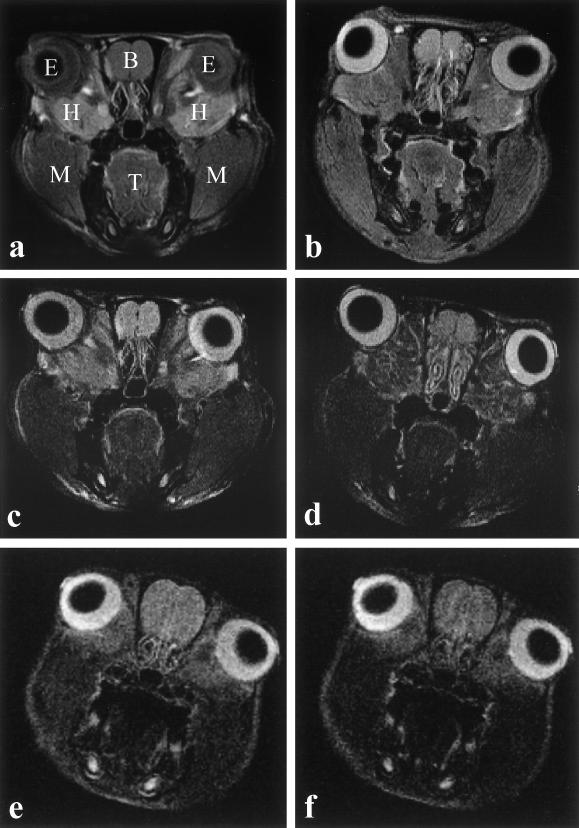
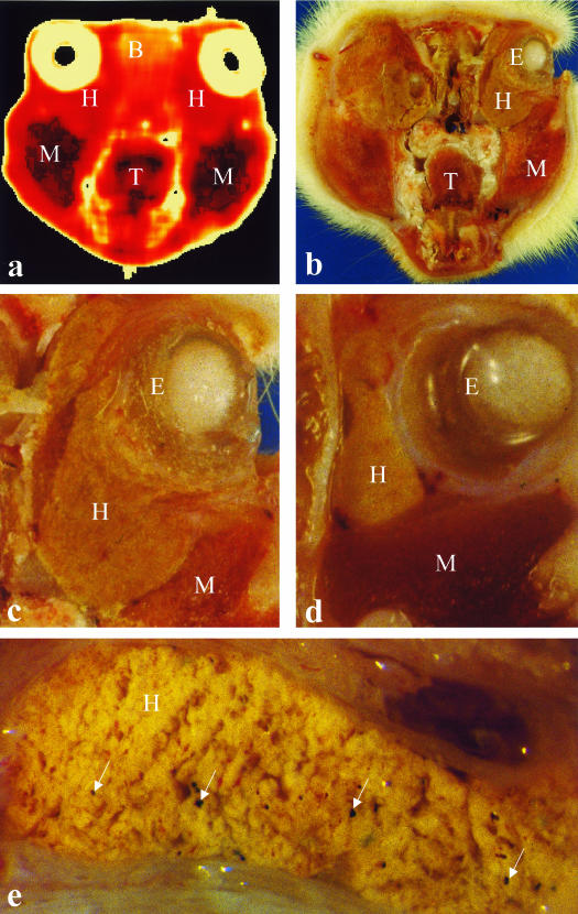
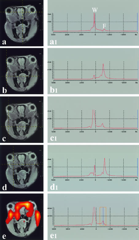
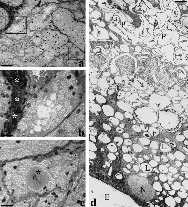
The intra-orbital lacrimal gland (Harderian gland, or HG) of the female rat was studied by magnetic resonance imaging (MRI) to evaluate whether MRI can be used to visualize the gland in vivo and localized-1H-spectroscopy detect its lipid content. The results were correlated with post-mortem anatomical sections, and with light and electron microscopy. On MRI, HG presented as a mass located between the ocular bulb and the orbit. In strongly T2W sequences the secretory structures had a reduced signal while intraparenchymal connective tissue was visible. T2-quantitative maps values of HG (60.12 +/- 8.15 ms, mean +/- SD) were different from other tissues (i.e. muscular tissue, T2 = 44.79 +/- 3.43 ms and olfactory bulb, T2 = 79.26 +/- 4.25 ms). In contrast-enhanced-MRI, HG had a signal-intensity-drop of 0.074 +/- 0.072 (mean +/- SD), after injection of AMI-25, significantly different from the muscle (0.17 +/- 0.10). Localized MRI spectra gave a large part of the signal originating from fat protons, but with a significant percentage from water protons. At light and electron microscopy the lipid deposition appeared to be composed of low-density material filling a large part of the cytoplasm, and the porphyrin aggregates were easily recognizable. The data demonstrate that an in vivo study of the HG was feasible and that high-field MRI allowed analysis of the gross anatomy detecting the lipid content of the gland.
Figures
Similar articles
-
Magnetic resonance imaging of the Harderian gland in piglets.J Anat. 2006 Nov;209(5):699-705. doi: 10.1111/j.1469-7580.2006.00642.x. J Anat. 2006. PMID: 17062026 Free PMC article.
-
Is it or isn't it? A reexamination of the anterior orbital glands of the fat-tailed Dunnart Sminthopsis Crassicaudata (Dasyuridae: Marsupiala) and a reevaluation of definitions for the Harderian gland.Anat Rec (Hoboken). 2010 Aug;293(8):1449-54. doi: 10.1002/ar.21111. Anat Rec (Hoboken). 2010. PMID: 20665822
-
The harderian gland and its excretory duct in the Wistar rat. A histological and ultrastructural study.J Anat. 1994 Jun;184 ( Pt 3)(Pt 3):553-66. J Anat. 1994. PMID: 7928644 Free PMC article.
-
Major ocular glands (harderian gland and lacrimal gland) of the musk shrew (Suncus murinus) with a review on the comparative anatomy and histology of the mammalian lacrimal glands.J Morphol. 1989 Jul;201(1):39-57. doi: 10.1002/jmor.1052010105. J Morphol. 1989. PMID: 2664187 Review.
-
Hormones and the control of porphyrin biosynthesis and structure in the hamster harderian gland.Microsc Res Tech. 1996 Jun 1;34(2):123-32. doi: 10.1002/(SICI)1097-0029(19960601)34:2<123::AID-JEMT5>3.0.CO;2-T. Microsc Res Tech. 1996. PMID: 8722706 Review.
Cited by
-
In vivo axonal transport deficits in a mouse model of fronto-temporal dementia.Neuroimage Clin. 2014 Mar 31;4:711-7. doi: 10.1016/j.nicl.2014.02.005. eCollection 2014. Neuroimage Clin. 2014. PMID: 24936422 Free PMC article.
-
Magnetic resonance imaging of orbital tumors.Eur Radiol. 2006 Oct;16(10):2207-19. doi: 10.1007/s00330-006-0227-0. Epub 2006 Apr 1. Eur Radiol. 2006. PMID: 16583212 Review.
-
[Appearance of orbital diseases on MRI].Radiologe. 2005 Sep;45(9):783-9. doi: 10.1007/s00117-005-1264-4. Radiologe. 2005. PMID: 16133401 Review. German.
-
Magnetic resonance imaging of the Harderian gland in piglets.J Anat. 2006 Nov;209(5):699-705. doi: 10.1111/j.1469-7580.2006.00642.x. J Anat. 2006. PMID: 17062026 Free PMC article.
-
Pharmocologic treatment with histone deacetylase 6 inhibitor (ACY-738) recovers Alzheimer's disease phenotype in amyloid precursor protein/presenilin 1 (APP/PS1) mice.Alzheimers Dement (N Y). 2015 Oct 11;1(3):170-181. doi: 10.1016/j.trci.2015.08.001. eCollection 2015 Nov. Alzheimers Dement (N Y). 2015. PMID: 29854936 Free PMC article.
References
-
- Antolin I, Rodriguez C, Uria H, Sainz RM, Mayo JC, Kotler ML, et al. Castration increases cell damage induced by porphyrins in the Harderian gland of male Syrian hamster. Necrosis and not apoptosis mediates the subsequent cell death. J. Struct. Biol. 1996;116:377–389. - PubMed
-
- Baccari GC. Organogenesis of the Harderian gland: a comparative survey. Microsc. Res. Techn. 1996;34:6–15. - PubMed
-
- Buzzell GR, Blank JL, Vaughan MK, Reiter RJ. Control of secretory lipid droplets in the Harderian gland by testosterone and the photoperiod: comparison of two species of hamsters. General Comp. Endocrinol. 1995;99:230–238. - PubMed
-
- Buzzell GR. Sexual dimorphism in the Harderian gland of the Syrian hamster is controlled and maintained by hormones, despite seasonal fluctuations in hormone levels: functional implications. Microsc. Res. Techn. 1996;34:133–138. - PubMed
-
- Cavagnari BM, Sterin-Speziale N, Affanni JM, Knudsen J, Santome JA. Acyl-CoA-binding protein in the armadillo Harderian gland: its primary structure and possible role in lipid secretion. Biochim. Biophys. Acta. 2001;1545:314–325. - PubMed
Publication types
MeSH terms
Substances
LinkOut - more resources
Full Text Sources
