. 2002 Sep;201(3):239-56.
doi: 10.1046/j.1469-7580.2002.00087.x.

The development of the gubernaculum and inguinal closure in the marsupial Macropus eugenii

Affiliations

The development of the gubernaculum and inguinal closure in the marsupial Macropus eugenii

Douglas Coveney et al. J Anat. 2002 Sep.

Abstract

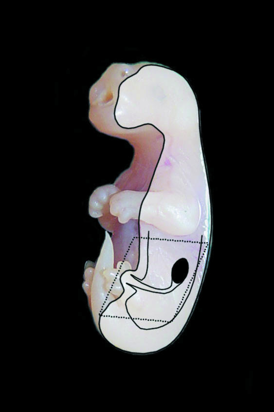
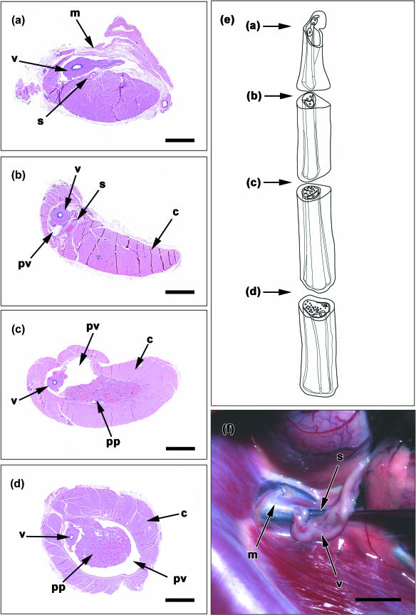
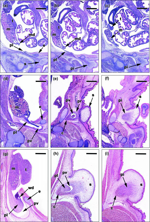
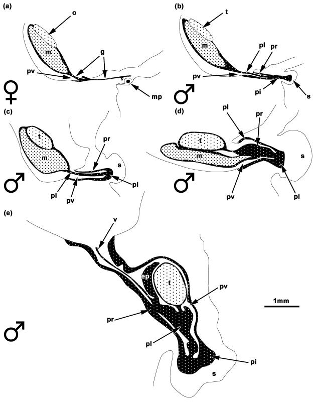
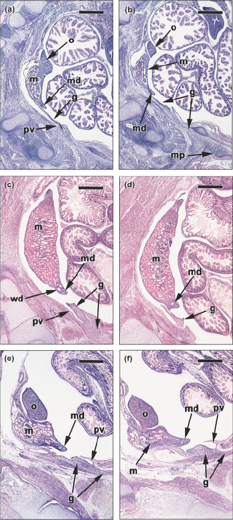
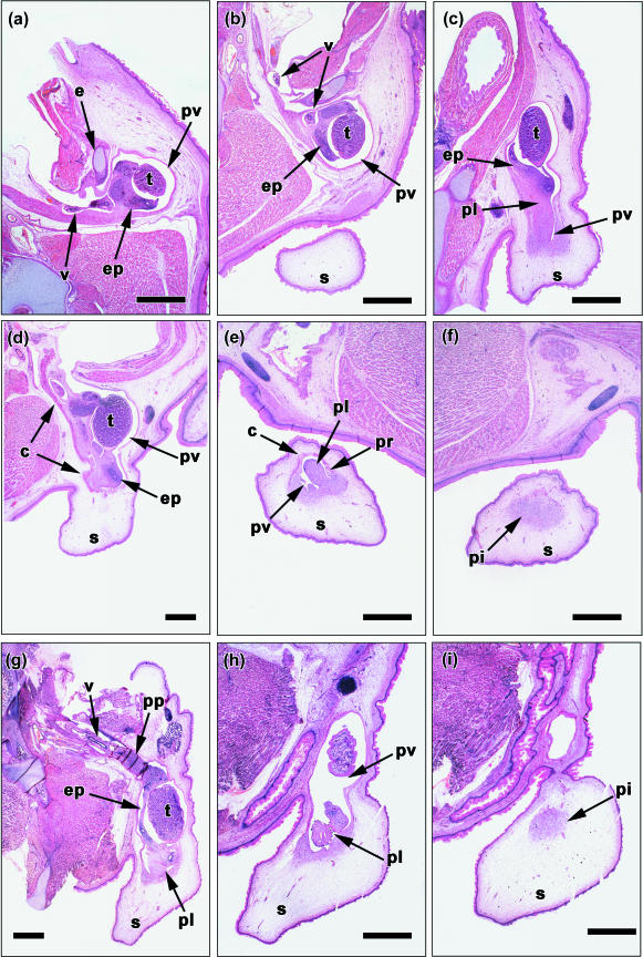
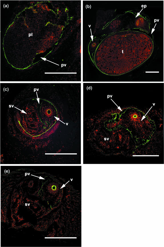
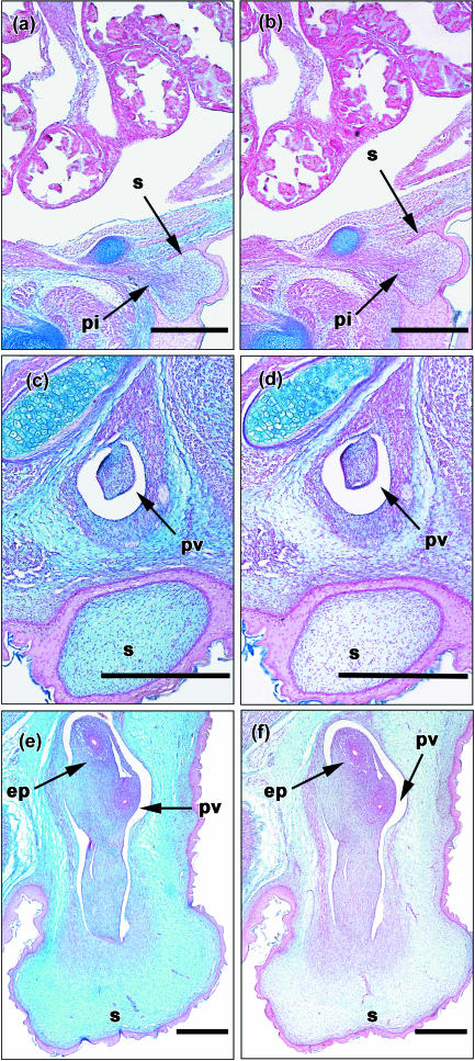
This study reports the developmental anatomy of testicular descent and inguinal closure of the tammar wallaby (Macropus eugenii) from birth to maturity. In females the ovary migrated caudally between days 10 and 20 after birth. The gubernaculum differentiates into the round ligament in the abdomen and extra-abdominally as the ilio-marsupialis muscle of the mammary glands. In males the testes migrated to the internal inguinal ring by day 20 post partum (pp), coinciding with the enlargement of the gubernaculum, and from the internal inguinal ring to the scrotum between days 20 and 65 pp. During descent there was an increase in the hyaluronic acid concentration in cells of the gubernaculum and scrotum. Development of the cremaster muscle began by day 10 pp on the periphery of the gubernaculum and its basic structure was completed by day 60 pp. After descent the inguinal canal closed between days 50 and 60 pp, but a small irregular lumen persisted, somewhat similar to that seen in the congenital scrotal hydrocoele of humans. Tammars have a hopping mode of locomotion and, like humans, are essentially bipedal. We suggest that inguinal closure evolved in these two species because their upright posture may otherwise lead to a high incidence of inguinal hernias.


Figures

Fig. 1
Fig. 1. The plane of histological sections in Figs 3, 5, 6 and 8 and in the two-dimensional reconstructions in Fig. 4. Sections were taken in a sagittal plane and the area of the gonad, mesonephros, gubernaculum and scrotum photographed.
Fig. 2
Fig. 2. The anatomy of the adult male inguinal canal. (a) A section of the inguinal canal at the level of the internal inguinal ring, showing the vas deferens (v) and spermatic vessels (s) lying underneath a membrane (m) that covers the entrance of the canal. (b) A section just distal to the inguinal ring, in which the thin processus vaginalis (pv) surrounds the vas deferens. The spermatic vessels are in a retro-peritoneal position and not within the processus vaginalis. The well developed cremaster muscle (c) runs along the dorsal aspect of the inguinal canal. (c) A section of the inguinal canal at a more posterior position, showing the vas deferens and pampiniform plexus (pp) situated in a larger processus vaginalis. (d) A section of an inguinal canal just proximal to the scrotum and testis, in which the vas deferens and large pampiniform plexus are situated within the processus vaginalis. At this level the cremaster muscle evenly surrounds the inguinal canal. (e) A schematic reconstruction of the adult inguinal canal showing the level of transverse sections represented in figures (a) to (d). (f) The internal inguinal ring of an adult male, showing the vas deferens and spermatic vessels entering the inguinal canal underneath a membranous flap that covers the internal inguinal ring. Scale bars represent 2 mm in a, b, c and d, and 5 mm in f.
Fig. 3
Fig. 3. A series of sagittal sections from lateral to medial (left to right) of male pouch young of the ages of day 0 (a–c), day 10 (d–f) and day 20 (g–i). (a) A lateral section of a day 0 male showing the testis (t) and mesonephros (m) situated high in the abdomen. The Müllerian duct (md) is situated at the posterior aspect of the mesonephros. A thin plica gubernaculi (pl) is present, surrounded by the processus vaginalis and pars vaginalis gubernaculi. The processus vaginalis teminates beside the epipubic bone (e). The scrotal bulges (s) are prominent. (b) A section showing the continuous gubernacular structure extending from the mesonephros to the scrotum. The plica gubernaculi is attached to the mesonephros and gonad and extends into the inguinal canal. The pars infravaginalis gubernaculi (pi) expands with the mesenchyme of the scrotum. (c) A medial section of a day 0 male showing the pars infravaginalis gubernaculi as a dark mass of mesenchyme within the scrotum. (d) A lateral section of a day 10 male showing the testis attached to the mesonphros situated within the abdominal cavity. The plica gubernaculi is attached to the mesophros and is surrounded by the processus vaginalis (pv). (e) A section showing the cremater muscle (c) developing on the dorsal aspect of the pars vaginalis gubernaculi (pr). The processus vaginalis is well developed and extends past the epipubic bone. (f) A medial section showing the pars infravaginalis gubernaculi within the scrotum. (g) A lateral section of a day 20 male in which the testis and regressing mesonephros are situated at the internal inguinal ring. The plica gubernaculi is larger than that of a day 10 male and contains the Wolffian duct (wd). (h) A section showing the processus vaginalis at the neck of the scrotum. The cremaster muscle is developing on the dorsal aspect of the pars vaginalis gubernaculi. (i) The pars infravaginalis gubernaculi is situated within the scrotum and the cremaster is developing on its dorsal aspect. Scale bars represent 500 µm.
Fig. 4
Fig. 4. Two-dimentional reconstructions of (a) a day 0 female, (b) a day 0 male, (c) a day 10 male, (d) a day 20 male, and (e) a day 30 male. Scale bar represents 1 mm. o = ovary; t = testis; m = mesonephros; g = gubernaculum; pl =plica gubernaculi; pr =pars vaginalis gubernaculi; pi =pars infravaginalis gubernaculi; pv = processus vaginalis; v = vas deferens; ep = epididymis; s = scrotum; mp = mammary primordium.
Fig. 5
Fig. 5. A series of sagittal sections from lateral to medial (left to right) of a female pouch young at day 0 (a,b), day 10 (c,d) and day 20 (e,f). (a) A lateral section of a day 0 female showing the ovary (o) and mesonephros (m) situated high in the abdomen. The gubernaculum (g) is attached to the posterior portion of the mesonephros, ovary and Müllerian duct (md) and extends into the abdominal wall. The processus vaginalis (pv) is present as a small cavity in the gubernaculum at the level of the abdominal wall. (b) A medial section of a day 0 female showing the gubernaculum extending from the abdominal wall to the dorsal aspect of the mammary primordia (mp). (c) A lateral section of a day 10 female showing the mesonephros, Müllerian and Wolffian ducts (wd) situated within the abdominal cavity. The gubernaculum extends past the abdominal wall, but the processus vaginalis teminates before the external inguinal ring. (d) A medial section of a day 20 female showing the attachment of the gubernaculum to the developing Müllerian duct. (e) A lateral section of a day 20 female, showing a well-developed ovary attached to a regressing mesonephros. The gubernaculum extends past the external inguinal ring, but the processus vaginalis remains small terminated within the inguinal canal. (f) A medial section of a day 20 female in which the gubernaculum is in close opposition with the Müllerian duct and extends past the abdominal musculature. Scale bars, 500 µm.
Fig. 6
Fig. 6. A series of sagittal sections from lateral to medial (left to right) of male pouch young at day 30 (a–c), day 40 (d–f) and day 50 (g–i). (a) A lateral section of a day 30 male in which the testis (t) is situated within the processus vaginalis (pv) just distal to the epipubic bone (e). The Wolfian duct is differentiating into the vas deferens (v) and epididymis (ep). (b) A section of a day 30 male showing the relative position of the testis and scrotum (s). (c) A medial section of a day 30 male showing the plica gubernaculi (pl) attached to the epididymis and extending into the scrotum. The processus vaginalis also terminates with the scrotum. (d) A lateral section of a day 40 male in which the well-developed testis is situated within the processus vaginalis. The cremaster muscle (c) is developing on the dorsal aspect of the inguinal canal. (e) The processus vaginalis, plica gubernaculi, pars vaginalis gubernaculi (pr) and the cremaster are present within the scrotum. (f) The pars infravaginalis gubernaculi is present with the scrotum situated distal to the processus vaginalis. (g) A lateral section of a day 50 male, in which the testis is situated at the neck of the scrotum. The vas deferens and pampiniform plexus (pp) are situated within the inguinal canal. The plica gubernaculi is attached to the epididymis and extends a short distance into the scrotum. (h) A section showing the processus vaginalis and plica gubernaculi with the scrotum.(i) The pars infravaginalis gubernaculi is present within the scrotum below the processus vaginalis. Scale bars, 1 mm.
Fig. 7
Fig. 7. Cytokeratin immunolocalization (green fluorescence) on male inguinal canals (transversely sectioned) which were counter stained with propidium iodide (red fluorescence) that stains nuclei. (a) The inguinal canal of a day 20 male in which cytokeratin was immunolocalized to epithelium of processus vaginalis (pv). The plica gubernaculi (pl) is situated within the processus vaginalis. (b) An inguinal canal from a day 40 male, in which the testis (t), epididymis (ep) and vas deferens (v) are surrounded by the processus vaginalis epithelium. (c) At day 50 the processus vaginalis epithelium surrounds most of the enlarged spermatic vessels (sv) and vas deferens. (d) An inguinal canal of a day 60 male, in which the processus vaginalis only surrounds the vas deferens. (e) An inguinal canal of a day 100 male showing the processus vaginalis epithelium persisting only arround the vas deferens. Scale bars, 500 µm.
Fig. 8
Fig. 8. Sagittal sectioned pelvic regions from male pouch young at days 0 (a,b), 10 (c,d) and 35 (e,f). (a) A control day 0 male stained with alcian blue, which stains glycosaminoglycans. Alcian blue staining is very weak in the gubernaculum (g), but stronger in the mesenchyme of the scrotum (s). (b) An adjacent section which has been incubated with hyaluronidase before alcian blue staining shows reduced staining in the scrotum and gubernaculum. (c) A control treated section from day 10 male showing strong alcian blue staining in both the gubernaculum and the scrotum, which is reduced by hyaluronidase treatment (d). (e) The strong alcian blue staining of both the gubernaculum and scrotum at day 35 is substantially reduced by hyaluronidase (f). Scale bars represent 500 µm. pv = processus vaginalis.

Similar articles

Cited by

References

    1. Alberts B, Bray D, Lewis J, Raff M, Roberts K, Watson JD. Molecular Biology of the Cell. 3rd edn. New York: Garland Publishing; 1994.
    1. Archer M, Clayton G. Vertebrate Zoology and Evolution in Australia. Carlisle, Western Australia: Hesperian Press; 1984.
    1. Atwell JD. Inguinal herniae and the testicular feminization syndrome in infancy and childhood. Br. J. Surg. 1961;49:367–371. - PubMed
    1. Atwell JD. Inguinal hernia in female infants and children. Br. J. Surg. 1962;50:294–297. - PubMed
    1. Backhouse KM, Butler H. The gubernaculum testis of the pig (Sus scropha) J. Anat. 1960;94:107–119. - PMC - PubMed

Publication types

LinkOut - more resources